

NNCI Image Contest 2026 - Unique
Most Unique Capability
This category celebrates the unique capabilities each site of the NNCI has on offer. Please check out the images below and read a little about the research behind them.
Vote Here
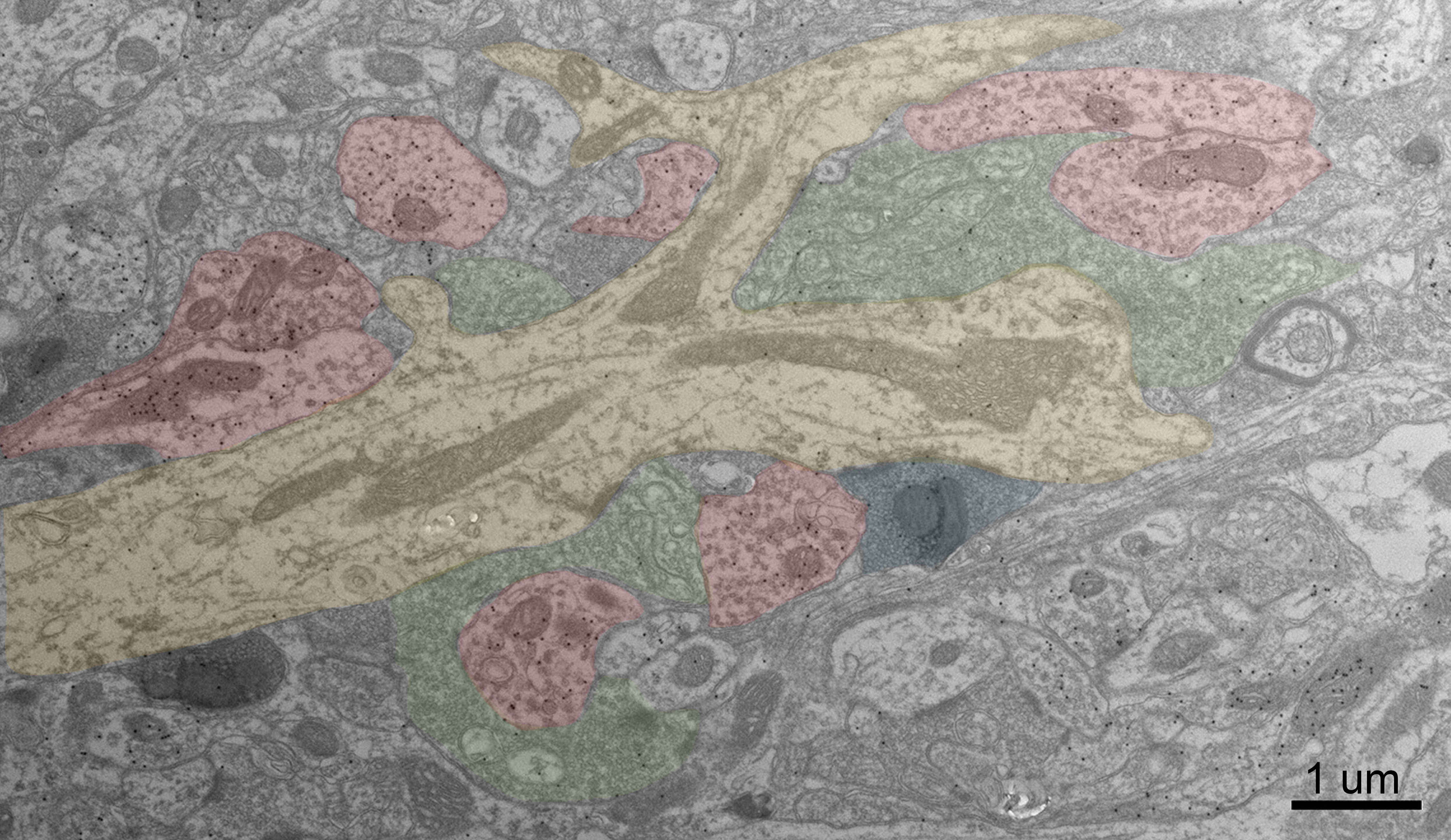
Convergence in the mouse thalamus
Artists: Kierra Mason (Graduate Student) & Martha Bickford (Professor), University of Louisville
NNCI Site: KY Multiscale
Tool: TEM - MNTC's BioEM Lab (UofL)
A transmission electron microscope image of the mouse dorsal lateral geniculate nucleus (dLGN). Synaptic terminals that converge to innervate a thalamocortical cell dendrite (pseudo colored yellow) are identified by viruses that express peroxidase in the cytoplasm (dark gray) or in mitochondria (pseudo colored blue), antibodies (pseudo colored red) or unique ultrastructure (pseudo colored green). Scale = 1 micron.
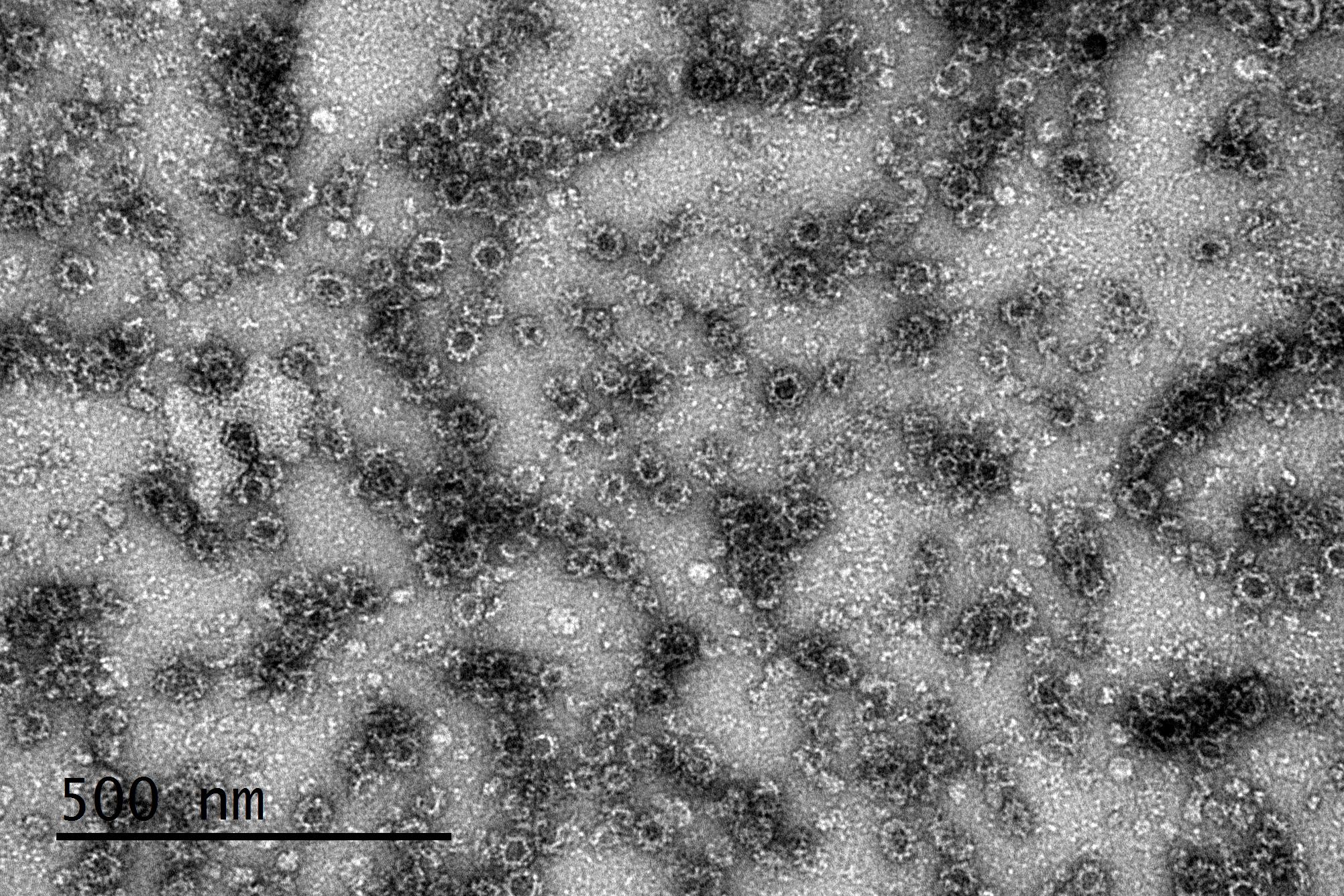
Human Norovirus GII.4 Viral Like Particles
Artists: Sai Navya Vadlamudi (Graduate Student) & Lijuan Yuan (Professor), Virginia Tech
NNCI Site: NanoEarth
Tool: JEOL JEM 1400
This transmission electron microscopy image reveals GII.4 human norovirus virus-like particles arranged in a highly organized, repeating T=3 capsid symmetry. Although these particles contain no viral genome and are non-infectious, they faithfully reproduce key structural features of the norovirus capsid. Their nanoscale organization provides a window into viral assembly and helps researchers understand how antibodies and other antiviral molecules recognize norovirus, knowledge that can contribute to the development of vaccines and antiviral strategies.
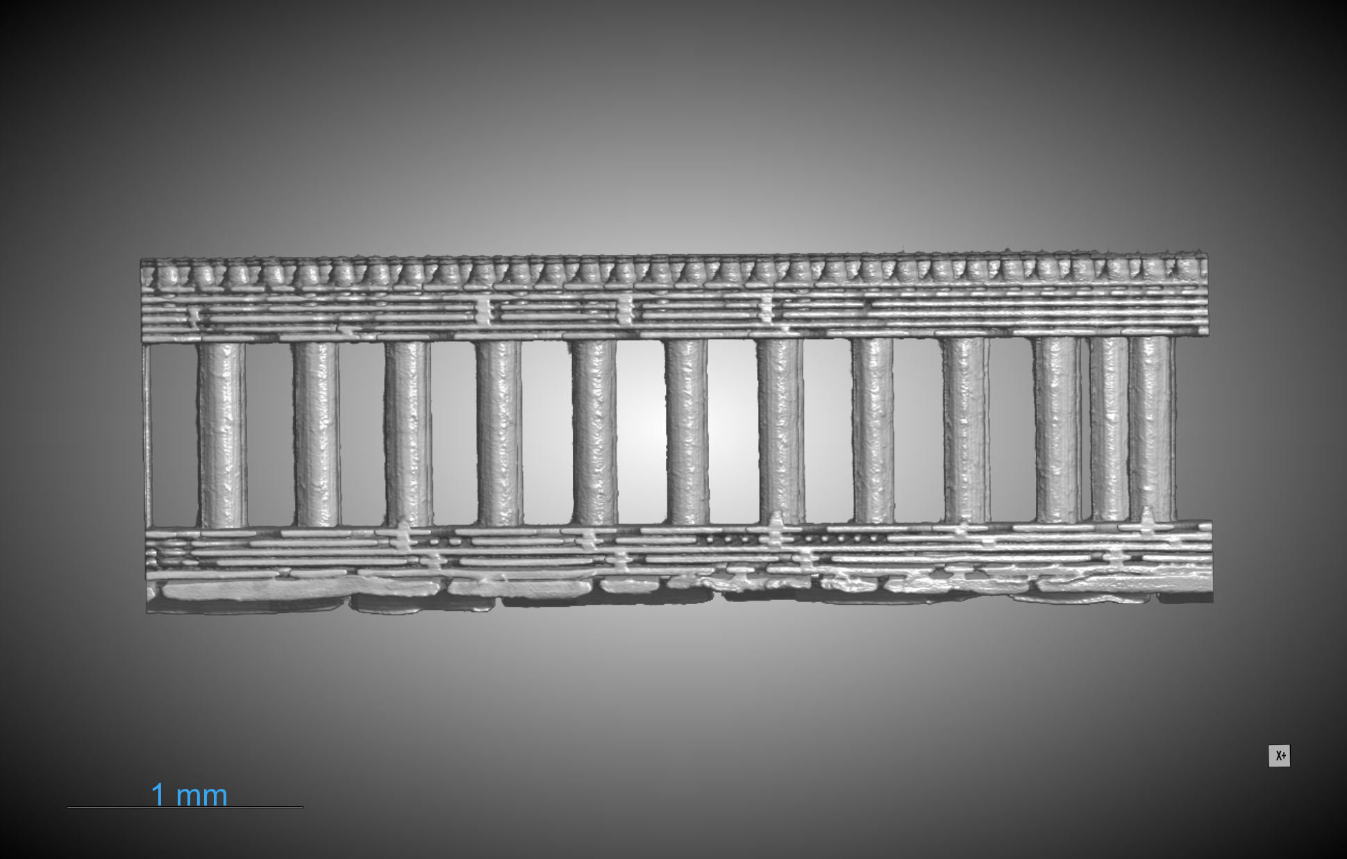
Inside Ryzen: The Architecture of Modern Computing
Artists: KyoungMok Kim (Graduate Student), Seung-Joon Paik (Research Engineer), Jack Moon (Research Scientist), Yongwon Lee (Research Engineer), Georgia Institute of Technology
NNCI Site: SENIC
Tool: Rigaku CT Lab HV
X-ray computed tomography reveals the internal architecture of a mechanically sectioned AMD Ryzen 5 7600X3D processor. Above the package interconnects, the reconstruction shows the stacked silicon dies behind AMD’s 3D V-Cache technology: a 64 MB SRAM cache die positioned above the Zen 4 Core Complex Die (CCD), which contains the processor cores and 32 MB of on-die L3 cache. The scan demonstrates CT’s ability to visualize complex three-dimensional semiconductor packaging structures, complementing destructive techniques such as SEM used to investigate the remaining section of the device.
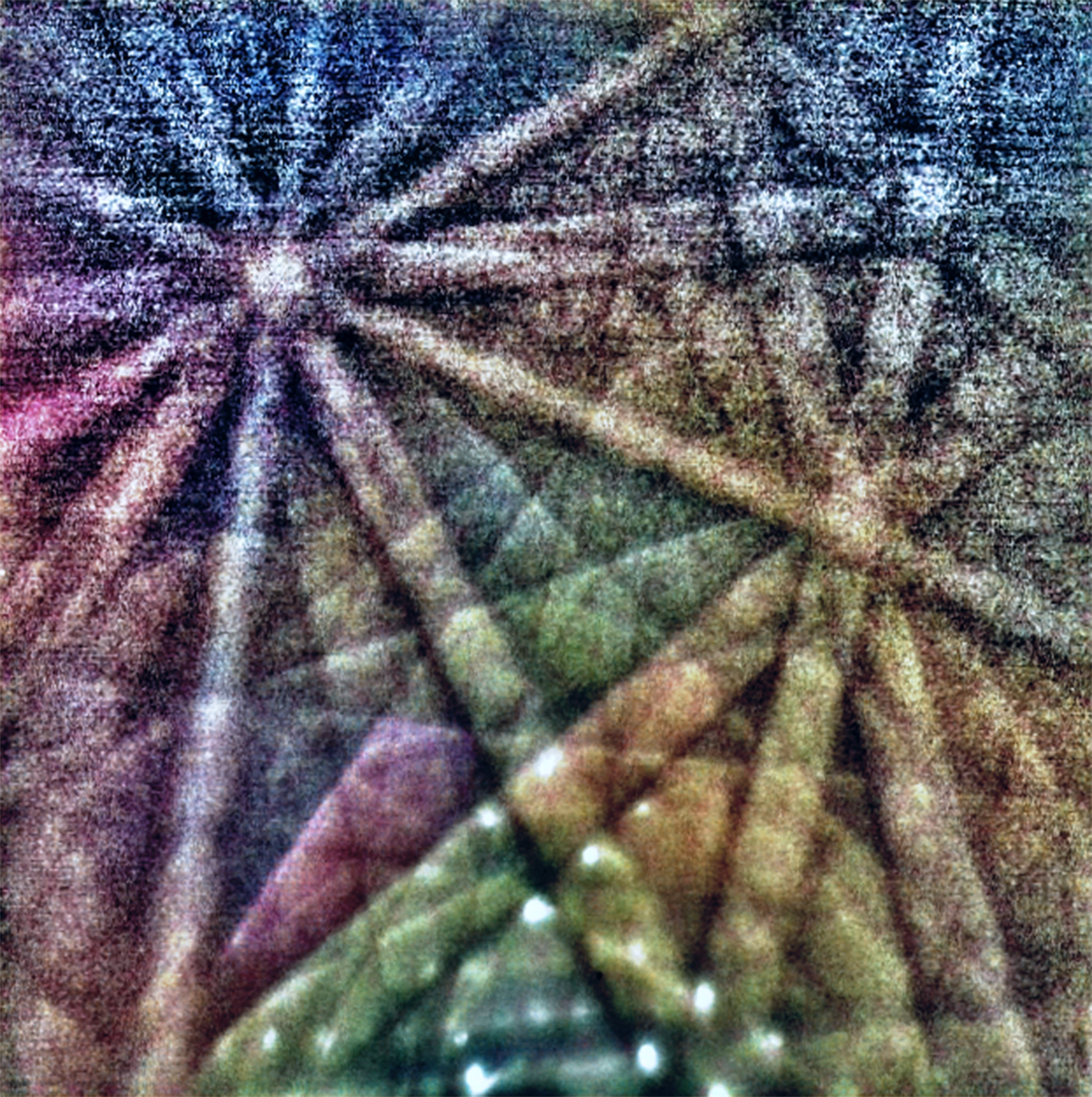
Star Atlas
Artist(s): Alp Kulaksizoglu (Graduate Student), Dravid Research Group, Northwestern University
NNCI Site: Northwestern University-NUANCE Center
Tool: Hitachi SU8700 SEM – TKD/EBSD pattern
Near-axis transmission kikuchi diffraction pattern of a nanoparticle featuring Kikuchi lines and Bragg spots (bottom middle) that look like stars.