
NNCI Image Contest 2025 - Whimsical
Most Whimsical
Artists in this category playfully used micro and nanoscale images as the foundation to build scenes. Please check out the images below and read a little about the research behind them.
Click Here to Vote
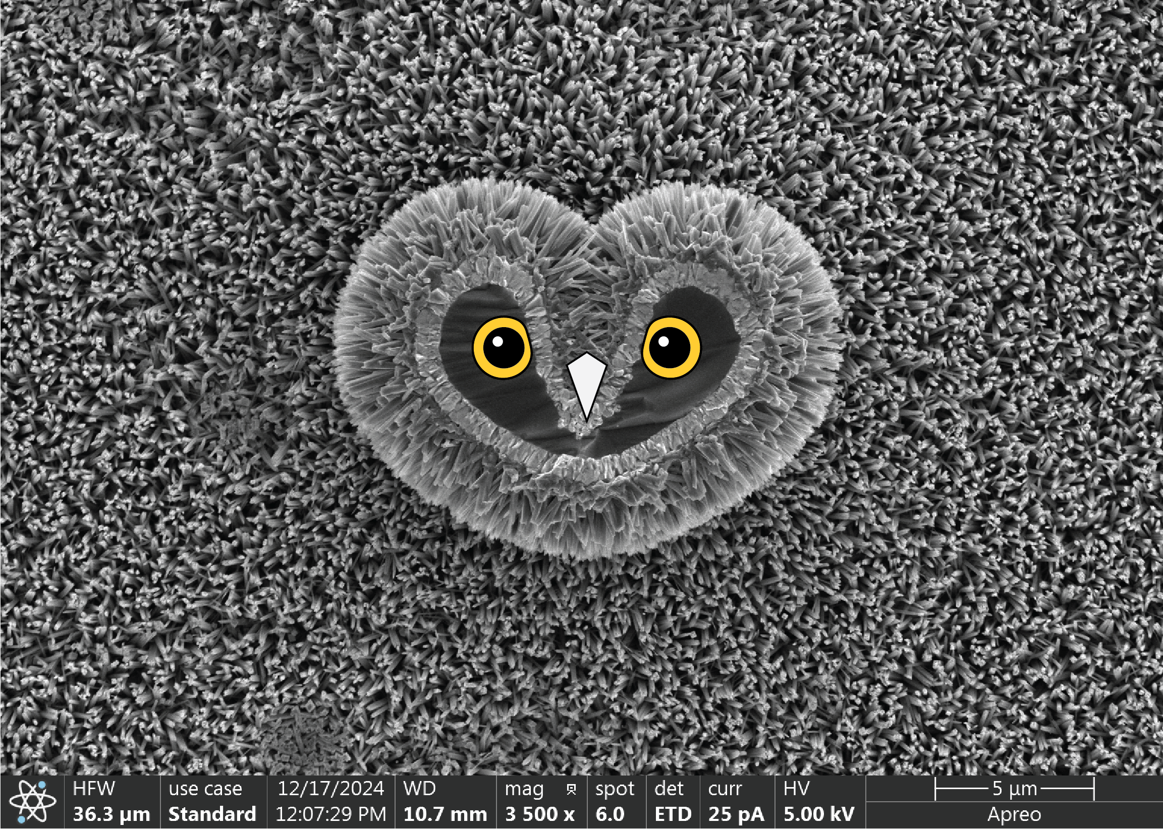
Love Owl
Artist: Matthew Mulvehill, Graduate Research Assistant, Conn Center for Renewable Energy Research, University of Louisville
NNCI Site: KY Multiscale
Tool: Thermo Fisher Apreo C LoVac Field Emission Scanning Electron Microscope (SEM) with high- and low-voltage ultra-high resolution capabilities, variable pressure, Back-scattered detector (BSD), Scanning Transmission Electron Microscopy (STEM) Detector and Energy-dispersive X-ray spectroscopy (EDS) Detector.
A scanning electron microscope (SEM) image of the left-over stump of a titanium dioxide nano-rod coated silicon micro-pillar that broke off from the substrate. The dark shadow around the owl’s eyes is silicon, the unique shape being an undesired artifact of a dry reactive ion etch (DRIE) during pillar formation. The silicon is coated with a thin layer of fluorine doped tin oxide followed by the titanium dioxide nano-rods that blanket the entire surface. The micro-pillar that was previously attached to the owl’s face will be used to sustainably generate hydrogen from only water and sunlight.
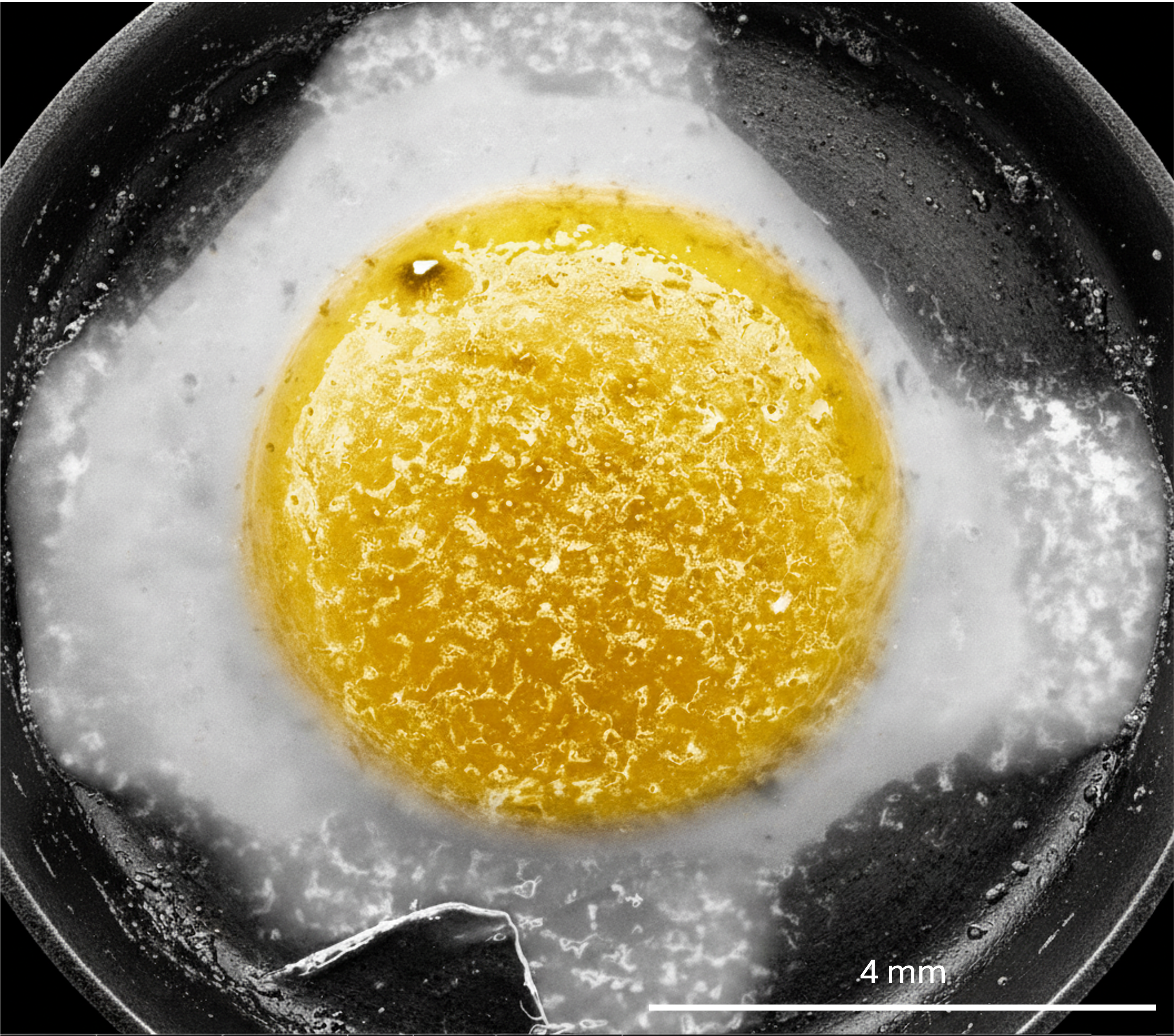
Sunny Side Up
Artist: Rayoon Kim, high school student, Engineering Summer Academy at the University of Pennsylvania
NNCI Site: MANTH
Tool: TFS Quanta 600 ESEM
At first glance, this might look like breakfast — but don’t grab a fork just yet. This is a scanning electron microscope image of a breathmint from Trader Joe’s, captured at 30 kV and later false-colored to highlight its egg-like features. The cracked central dome resembles a bright yolk, while the surrounding fractured shell mimics a sizzling egg white. This image shows that even everyday materials like breathmints can reveal porous textures at the microscale. It’s a reminder that science can be both serious and sunny side up.
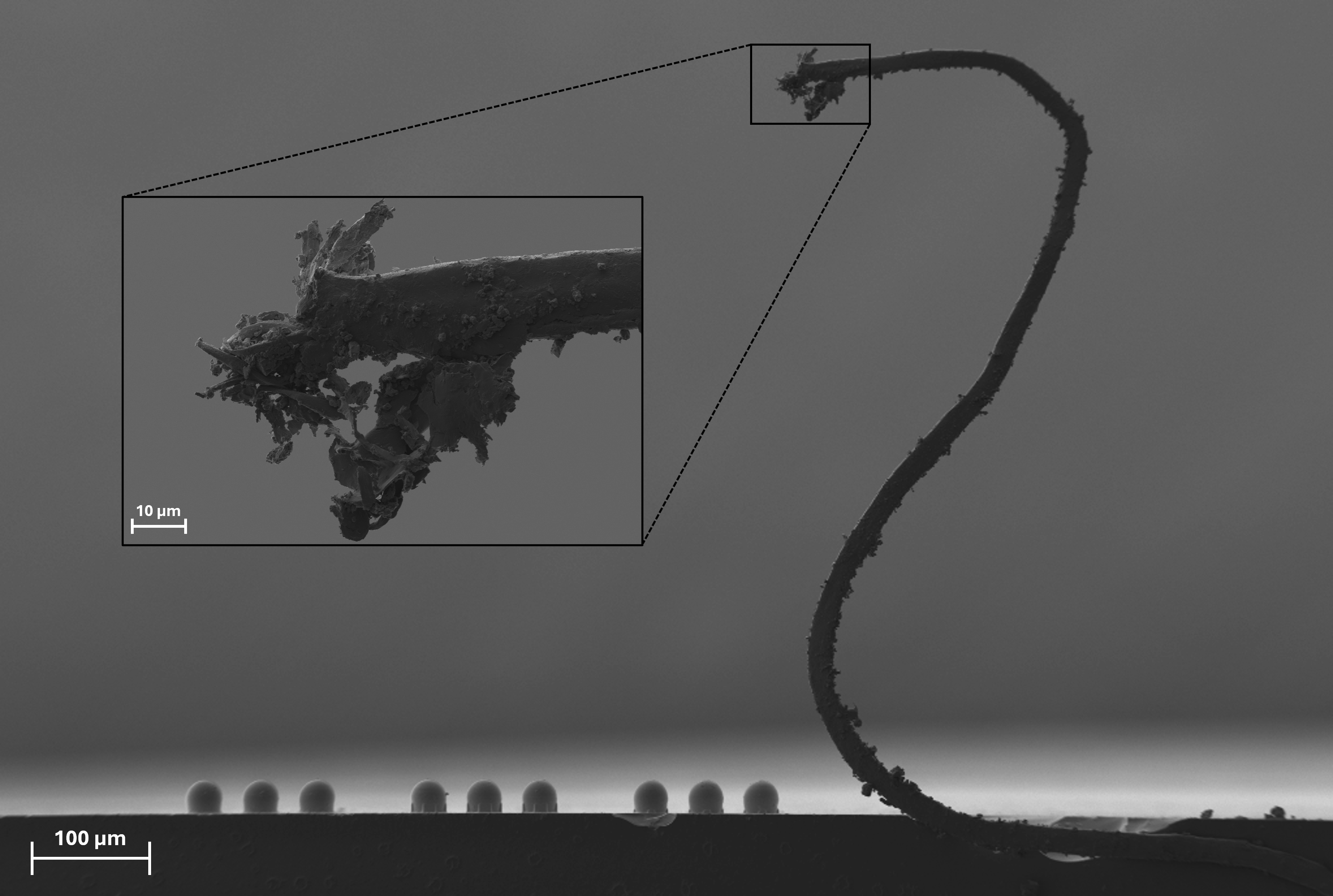
The Leviathan
Artists: Jordan Baker (Graduate Student, Nano-Optics Group), and Corey Pearson (Graduate Student, Spectrum Lab), Montana State University
NNCI Site: MONT
Tool: Zeiss SUPRA 55VP FE-SEM
The infamous Leviathan rears tall above an array of 3D-printed microspheres in an otherwise desolate scene. The inset image reveals that the head of the Leviathan is shockingly detailed, including a gaping maw with teeth and a curled tongue, a nose, eyes, and eyebrows. Like the Leviathan itself, this structure was unlikely and unexpected; it is likely of a stray strand of iridium-coated carbon tape that was adhered to the glass substrate. Different support structures were tested with the microspheres since both diameter and fidelity to spherical shape may allow these printed devices to emulate optical scattering through fog.
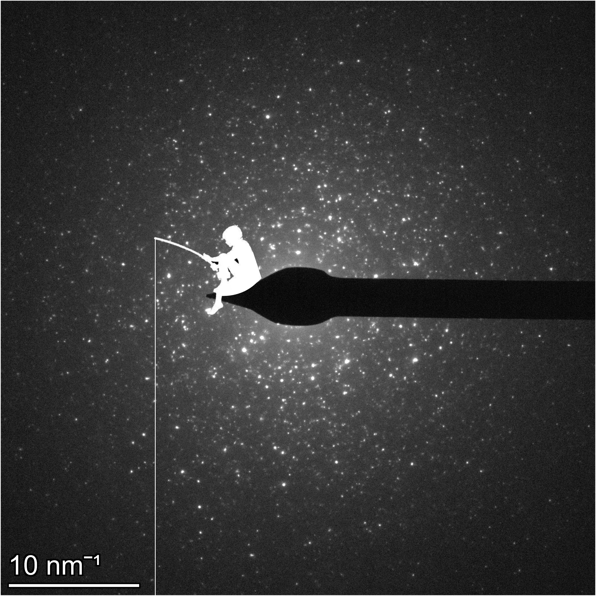
NanoDreamWorks
Artist: Abigail Carbone, Graduate Student, Stanford University
NNCI Site: nano@stanford
Tool: Thermo Fisher Spectra 300 TEM
It may seem that the stars millions of light years away and the atoms at our fingertips are worlds apart, but high-resolution electron microscope imaging suggests otherwise-- this image is a diffraction pattern of indium oxide, showcasing the beautiful, chaotic, and perhaps familiar nature of atomic structures. Since this image bears a striking resemblance to the night sky, I figured the boy sitting on the moon from the DreamWorks logo would feel right at home perched on top of the beam stop.
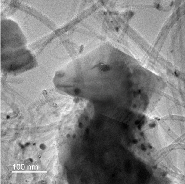
Little Lamb in the Lattice
Artist: Md Sifat Hossain, PhD student, Department of Chemical Engineering, Virginia Tech
NNCI Site: NanoEarth
Tool: JEOL JEM-2100 TEM
In this whimsical entry, a TEM image of a used catalyst reveals an unexpected surprise — the soft silhouette of a lamb. What you see is carbon deposition on the zeolite surface after methane conversion, with tiny carbon nanotubes creating intricate textures. While this image captures important details about catalyst deactivation, its playful resemblance to a lamb highlights the beauty of science beyond data and numbers. It reminds us that nanoscale research often unveils patterns and forms that spark imagination, offering a unique bridge between rigorous materials science and artistic interpretation at the atomic scale.
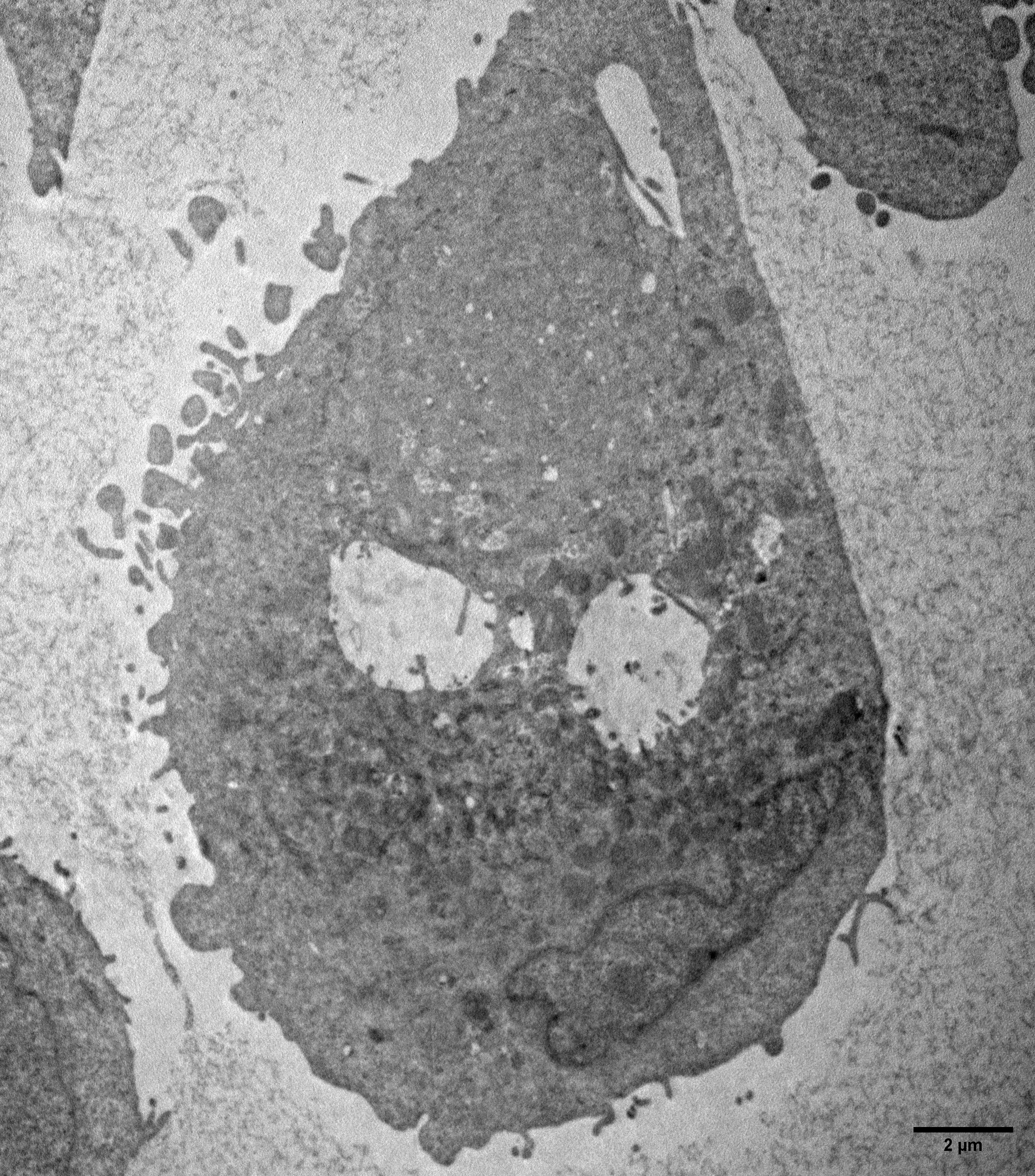
Cancer Can Be Such a Grinch
Artists: Portia N.A. Plange (graduate student) and Nicole M. Iverson (Associate Professor), University of Nebraska-Lincoln
NNCI Site: NNF
Tool: Transmission Electron Microscopy (TEM)
This image shows a breast cancer cell post incubation with Single Walled Carbon Nanotubes. The cancer cell looks like a 'bad guy' – which it is when it comes to human health.
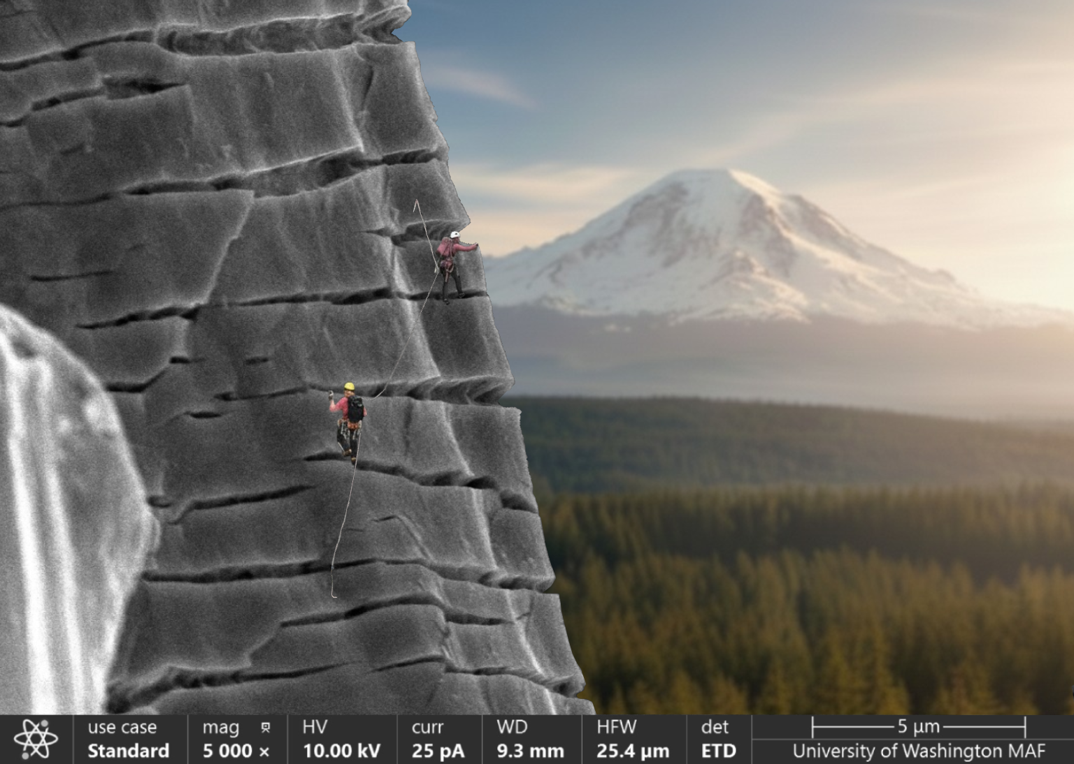
From Cracks to Summits (Don’t Worry, It’s Safe to Touch)
Artists: Jeongsu Shin (Graduate Student, University of Washington, LAMP Arola Group) and Dwayne D. Arola (Professor, University of Washington, LAMP Arola Group)
NNCI Site: NNI
Tool: ThermoFisher Scientific Apreo-S SEM
This image shows the pore surface of a titanium alloy sample (Ti-6Al-4V) manufactured by additive manufacturing after a tensile test. Normally the surface is smooth, but the pulling force causes cracks that look like tiny climbing routes on a rock wall. To highlight this, we imagined climbers scaling these microscopic cracks with Mount Rainier in the background. The scene reflects the spirit of research like climbers, we ascend together, overcoming obstacles and reaching for greater heights. The picture was captured at the University of Washington’s Molecular Analysis Facility using an Apreo-2S scanning electron microscope.
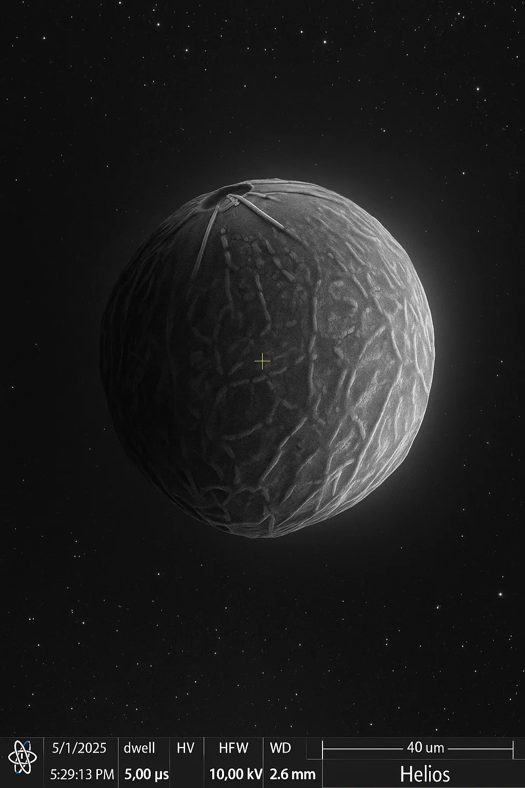
That’s No Moon
Artists: Justin Nakamura, PhD Student, Georgia Institute of Technology
NNCI Site: SENIC
Tool: FEI Helios FIB-SEM
This is a hydrogen generating nanoparticle that when exposed to any liquid with water (wastewater, urine, blood) it rapidly generates hydrogen for capture. Manufactured in-house at the Georgia Tech Advanced Manufacturing Pilot Facility (AMPF), this research aims to provide a cheaper manufacturing method to generate spherical hydrogen-generating nanoparticles 24/7 with limited human interaction.
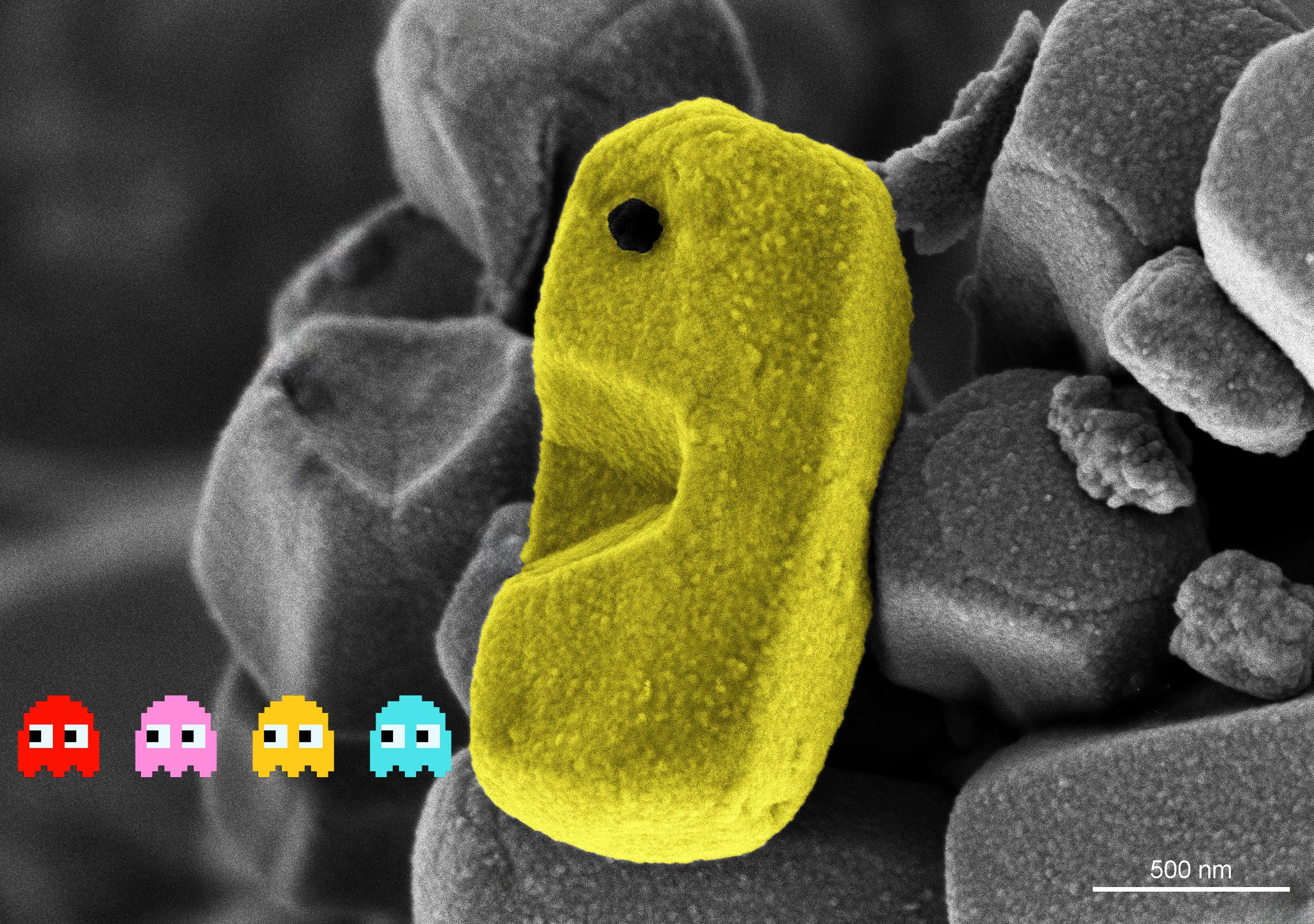
PAC-MOF
Artist: Nick Gogola, MS, Assistant Core Scientist, NUANCE Center / SHyNE Resource, Northwestern University
NNCI Site: SHyNE
Tool: Hitachi Hitachi SU8700 SEM
This SEM image shows an NU-1000 metal organic framework (MOF) crystal, displaying its characteristic faceted morphology. The colorization and graphics reimagine this crystal as an oversized PACMAN turning the tables on the persistent ghosts Inky, Blinky, Pinky, and Clyde. MOFs are porous materials composed of metal nodes and organic linkers which are widely studied for applications including catalysis, water purification, and carbon capture. This research aims to further study the morphology of the MOF crystals as this aspect is directly tied to their performance in these key areas.
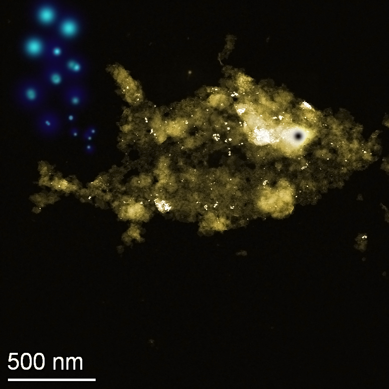
Nano - Au (Gold) Fish
Artist: Rishi Raj, PhD student, Department of Chemical Engineering and Material Science, University of Minnesota
NNCI Site: MINIC
Tool: ThermoFisher Talos F200X
The image shows a HAADF-STEM acquisition of a Au nanoparticle agglomerate that appears to have similar likeness as that of a fish, hence a nano-goldfish. These are Au nanoparticles used for various applications which were being studied by me for their quality, density, size and distribution along with a morphological understanding. The image was recolored to appear as gold contrast and cartoonistic bubbles were added for artistic effect