
NNCI Image Contest 2025 - Unique
Most Unique Capability
This category celebrates the unique capabilities each site of the NNCI has on offer. Please check out the images below and read a little about the research behind them.
Click Here to Vote
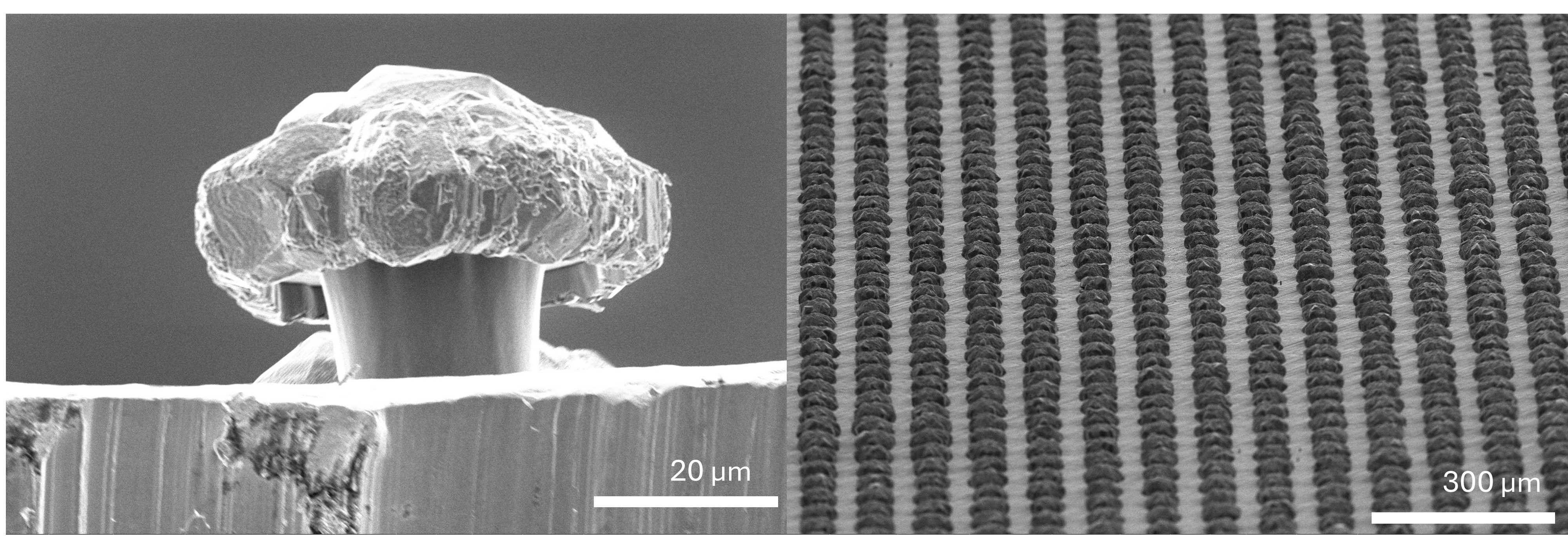
Micro-Mushroom Array
Artist: Hyogyun Roh, Post Doctoral Fellow, Conn Center for Renewable Energy Research, University of Louisville
NNCI Site: KY Multiscale
Tool: Thermo Fisher Apreo C LoVac Field Emission Scanning Electron Microscope (SEM) with high- and low-voltage ultra-high resolution capabilities, variable pressure, Back-scattered detector (BSD), Scanning Transmission Electron Microscopy (STEM) Detector and Energy-dispersive X-ray spectroscopy (EDS) Detector.
tion (100 words): Fabricated "doubly-re-entrant structured Cu mushroom array”. This material was used to create a superhydrophobic surface. The images show the doubly re-entrant structure of micromushrooms in the underside view (left) and uniform mushroom array in the top view (right).
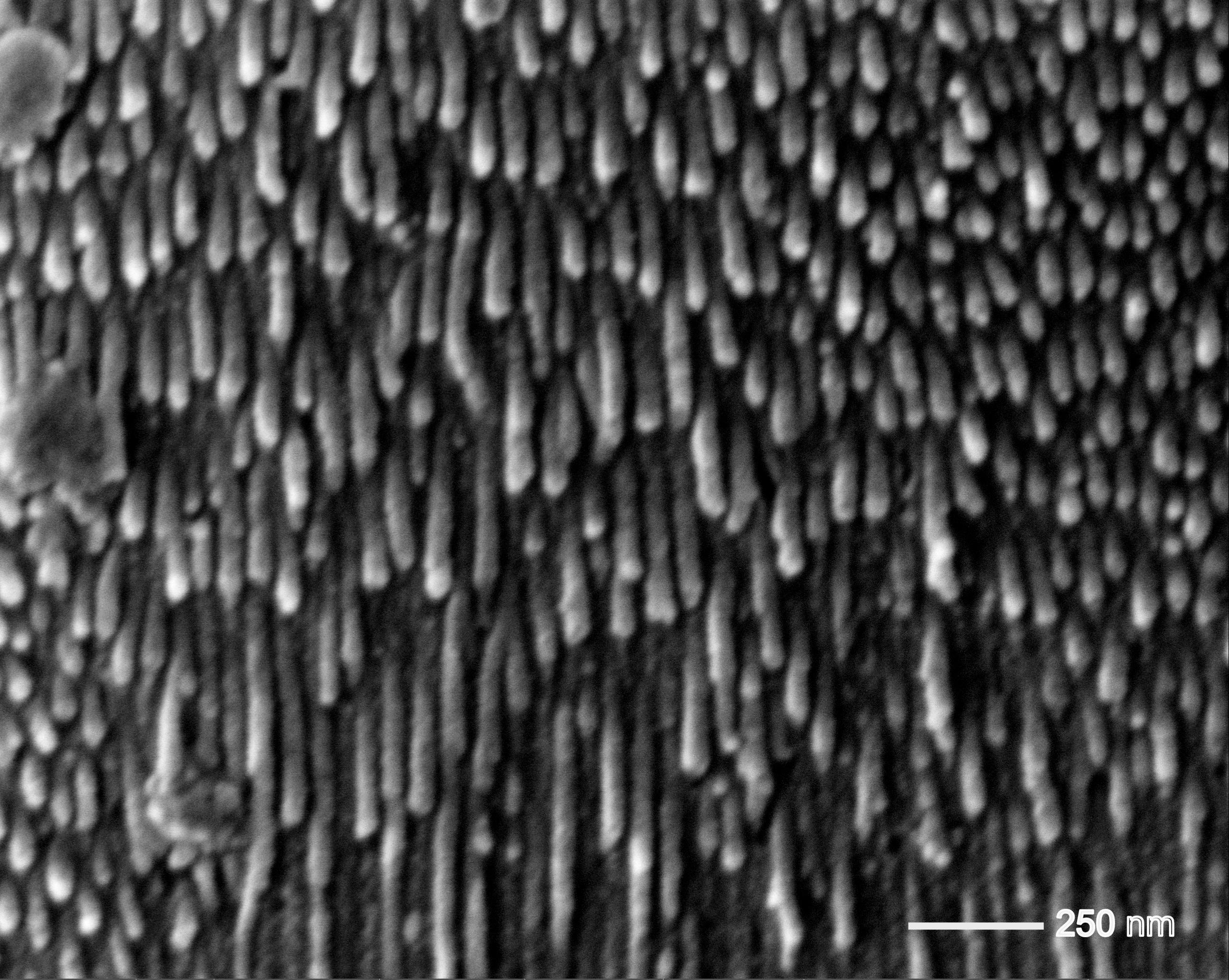
Meteor Shower in a Nanoworld
Artist: Sadikshya Pandey, PhD student, Department of Chemical Engineering and Material Science, University of Minnesota
NNCI Site: MINIC
Tool: Thermo Apreo.
This image shows the cross-section of porous alumina, created by anodizing aluminum in an acidic electrolyte. The process produces ordered arrangement of nanoscale pores that extend deep into the material, forming a structure invisible to the naked eye. Under the scanning electron microscope, these vertical channels resemble streaks of light, resembling a meteor shower in the night sky.
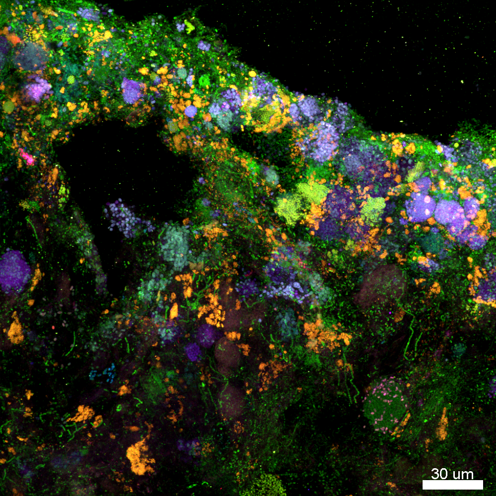
Lighting up the hidden life of wastewater bacteria
Artist: Kylie Bodle, postdoctoral researcher, Center for Biofilm Engineering, Montana State University
NNCI Site: MONT
Tool: Inverted Leica DMI8 Stellaris confocal laser scanning microscope
This image shows four bacterial groups in a wastewater biofilm: green indicates all bacteria, blue marks phosphate-accumulating organisms, red shows ammonia-oxidizing bacteria, and purple highlights glycogen-accumulating organisms. Each target was labeled using DNA strands tagged with fluorescent dyes, which stain only organisms containing the complementary DNA. While most confocal microscopes separate only red, green, and blue signals, the CBE’s microscope uses a white light laser, enabling detection of additional targets. This produces purple-colored cells and demonstrates the microscope’s unique imaging capability.
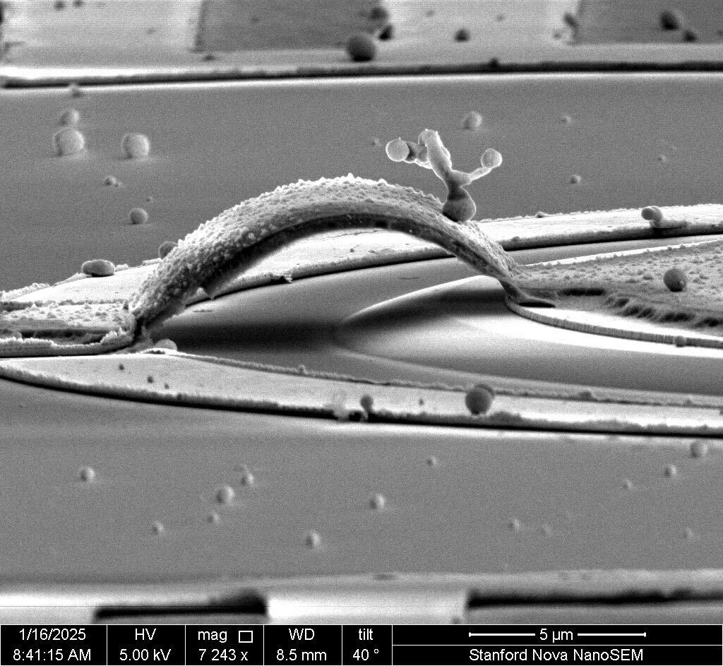
Tree on a Bridge
Artist: Dominic Catanzaro, Graduate Student, Stanford University
NNCI Site: nano@stanford
Tool: Nova SEM
This SEM showcases a carefully engineered airbridge upon which a golden tree has spontaneously grown, showcasing the beauty in both engineering and nature. During the testing of these structures, the explosive demise of one bridge lead to the growth of this beautiful tree upon the bridge pictured here. The adjacent bridge's demise is a scientific setback, but the growth of this tree is a reminder that in death there is life, even at the nanoscale.
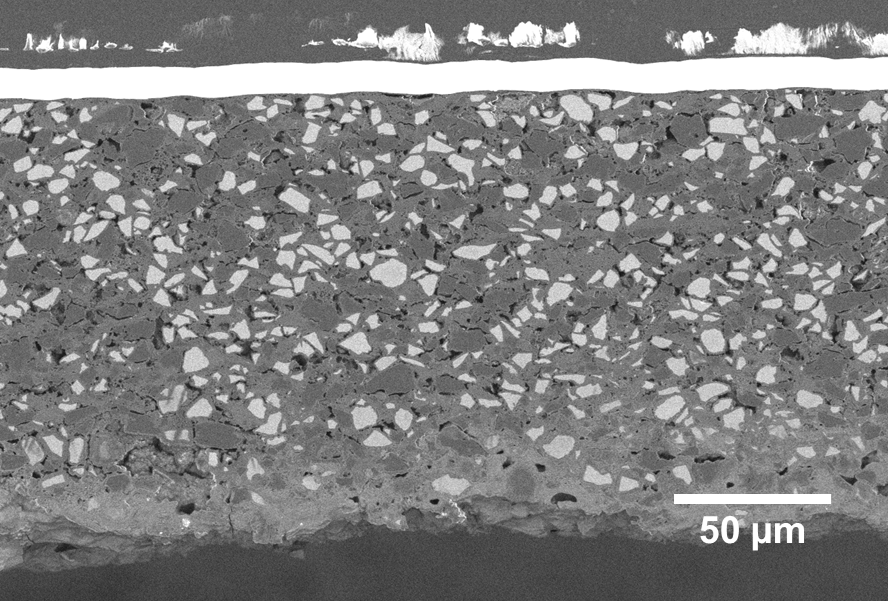
Advancing Post-Mortem Analysis: Wide-Area Cooling Cross-Section Polishing of a 1 Ah Pouch Cell Anode
Artists: Rupayan Ghosh (PhD Candidate, Department of Chemistry) and Jarret Wright (FIB/SEM Laboratory Manager, Nanoscale Characterization and Fabrication Laboratory), Virginia Tech
NNCI Site: NanoEarth
Tool: JEOL IT-500HR and JEOL IB-19520CCP Cooling Cross Section Polisher
This BSE image shows the wide-area cross-section of a 1Ah pouch cell SiOx@C composite anode, casted on Cu foil after long-term cycling. The sample was polished using the JEOL IB-19520CCP Cooling Cross Section Polisher and imaged using the JEOL IT-500HR, revealing the intricate internal morphology of the composite anode following extensive charge–discharge cycles.
Our group focuses on the synthesis and investigation of electrode materials under diverse cycling conditions through systematic post-mortem and teardown studies. With NCFL’s advanced cooling cross-section polisher, we can achieve wide-area cross-sectioning, providing comprehensive statistical insights that accelerate the understanding and development of next-generation, durable energy storage systems.
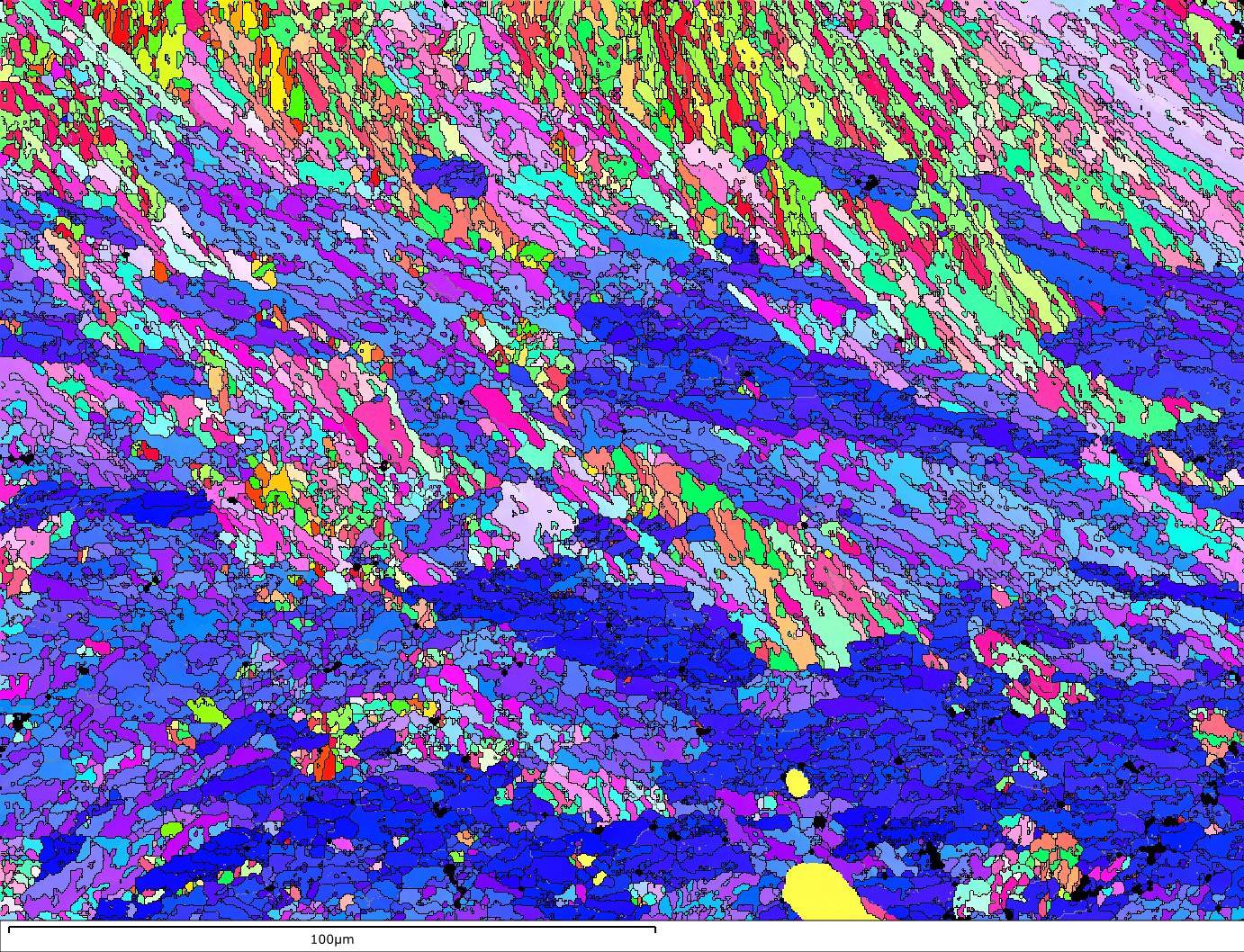
Waves in Stone
Artist: Yu Wen, PhD, Postdoctoral Researcher, NUANCE Center / SHyNE Resource, Northwestern University
NNCI Site: SHyNE
Tool: Hydra Plasma FIB
An electron backscatter diffraction map of coral’s aragonite skeleton reveals crystalline waves frozen in time. Blue bands mark grains aligned along the ⟨001⟩ axis, while greens and reds trace ⟨100⟩ and ⟨010⟩. Together, they form a vivid seascape where mineral order meets the rhythm of the ocean.
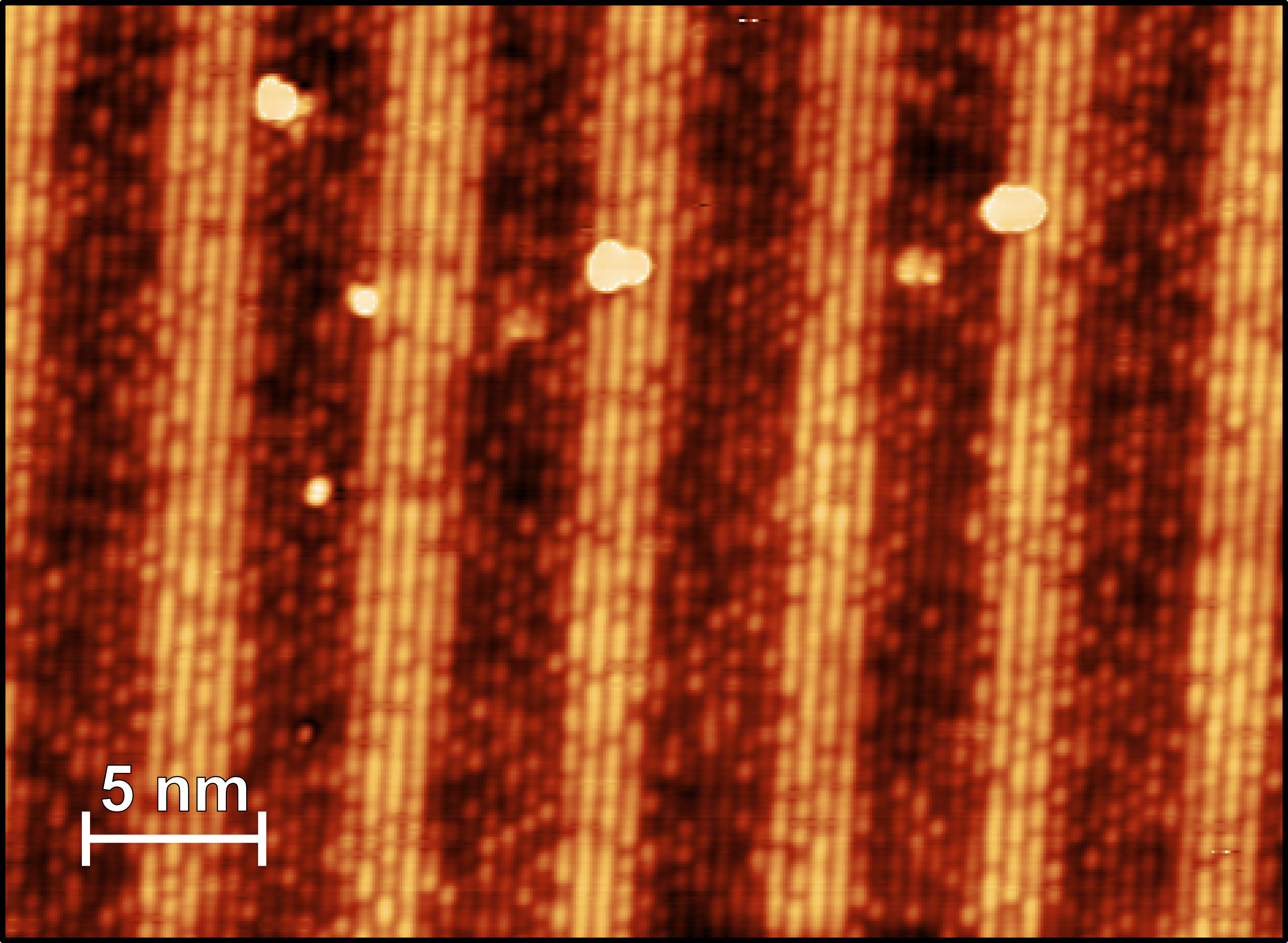
InAsSb Atomic Cross Section
Artist: Isaiah Ertel, Graduate Student, School of Physics, Georgia Institute of Technology
NNCI Site: SENIC
Tool: Createc LT-SPM
Shown here is an atomically resolved cross-sectional image of a layered semiconductor material. The sample was cleaved in ultrahigh vacuum and imaged with scanning tunneling microscopy at 77 K. The alternating bright and dark bands correspond to four atomic layers of InAs0.235Sb0.765 (bright) followed by seven atomic layers of InAs0.665Sb0.335 (dark). Within each band, the finer periodic corrugations reflect the atomic-scale lattice of the constituent layers.