
NNCI Image Contest 2025 - Stunning
Most Stunning
This category celebrates the beauty of the micro and nanoscale. Please check out the images below and read a little about the research behind them.
Click Here to Vote
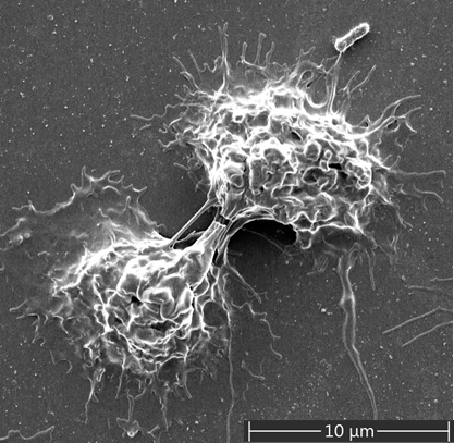
Guardians in Conversation
Artist: Shikha Srivastava, Graduate Research Assistant, University of Louisville.
NNCI Site: KY Multiscale
Tool: Thermo Fisher Apreo C LoVac Field Emission Scanning Electron Microscope (SEM) with high- and low-voltage ultra-high resolution capabilities, variable pressure, Back-scattered detector (BSD), Scanning Transmission Electron Microscopy (STEM) Detector and Energy-dispersive X-ray spectroscopy (EDS) Detector.
Two neutrophils, the body’s first defenders, connect with each other as if quietly communicating during an infection. This small interaction shows how they work together, turning a tiny moment into a powerful display of teamwork and defense.
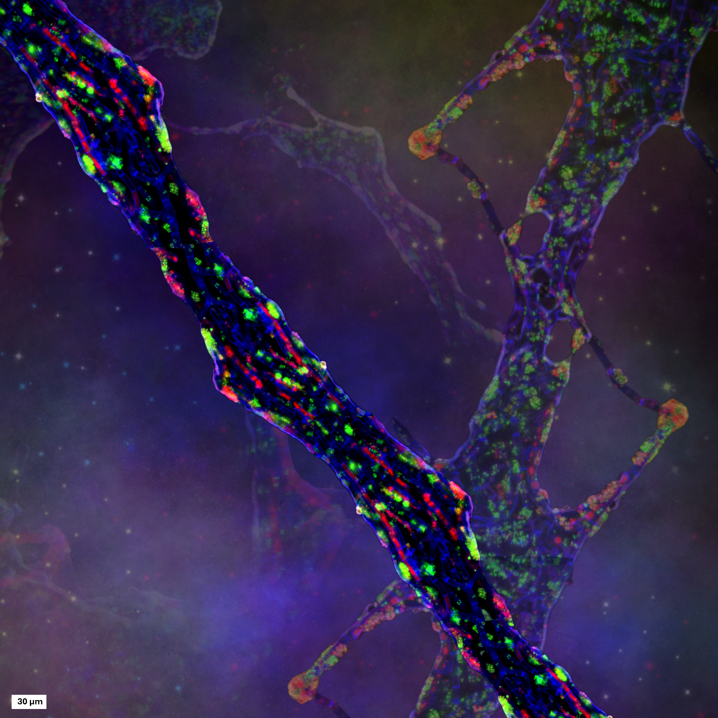
The Microbial Milky Way
Artist: Ghazal Vahidi, postdoctoral researcher, Center for Biofilm Engineering, Montana State University
NNCI Site: MONT
Tool: Leica Thunder Widefield Imager
This image reimagines a cross-kingdom biofilm as a cosmic scene. Blue fungal filaments (A. aculeatus) stretch like cosmic arms, while clusters of P.aeruginosa (green) and other DNA-rich material glow like stars. The composition was created by overlaying and stitching microscopy images onto a cosmic background. In reality, these images highlight a multi-domain biofilm, an ecosystem where organisms from different domains of life coexist and interact at the microscopic scale within a dense microbial communities embedded in protective extracellular material. What resembles a starry sky is in fact a vibrant microbial community, showcasing life building beautiful shared spaces between unlikely residents.
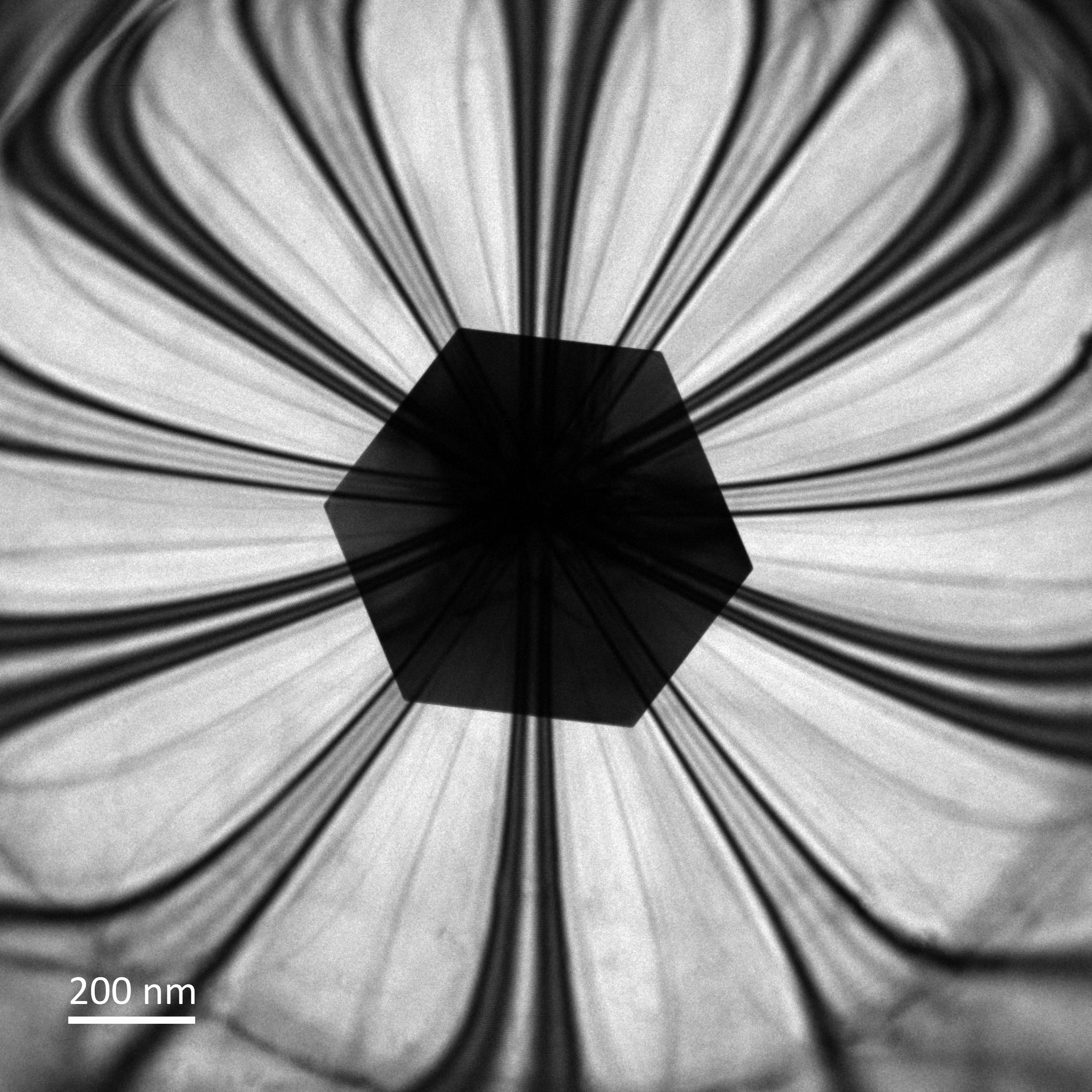
Twisted Cu Flower
Artist: Pawel Czaja, Post Doctoral Scholar, Stanford University
NNCI Site: nano@stanford
Tool: FEI Titan Environmental Transmission Electron Microscope 300 kV
The image shows a copper single crystal that has grown between two sheets of molybdenum disulfide (MoS₂). These two MoS₂ layers are not perfectly aligned: the top one is slightly rotated compared to the bottom one by a set “twist angle.”
At first, when deposited on bottom substrate with no capping layer the copper crystal grows epitaxially i.e. in line with the bottom MoS₂ layer, almost like it is copying its pattern. However, when the sample is capped with the top layer and heated to about 600°C, the crystal starts to adjust its orientation towards the top layer. As a result, the copper crystal slowly shifts its orientation so that it is no longer matching only the bottom layer but instead settles into a position in between the two — a compromise that balances the pull of both layers, demonstrating a recently discovered phenomenon called "twisted epitaxy".
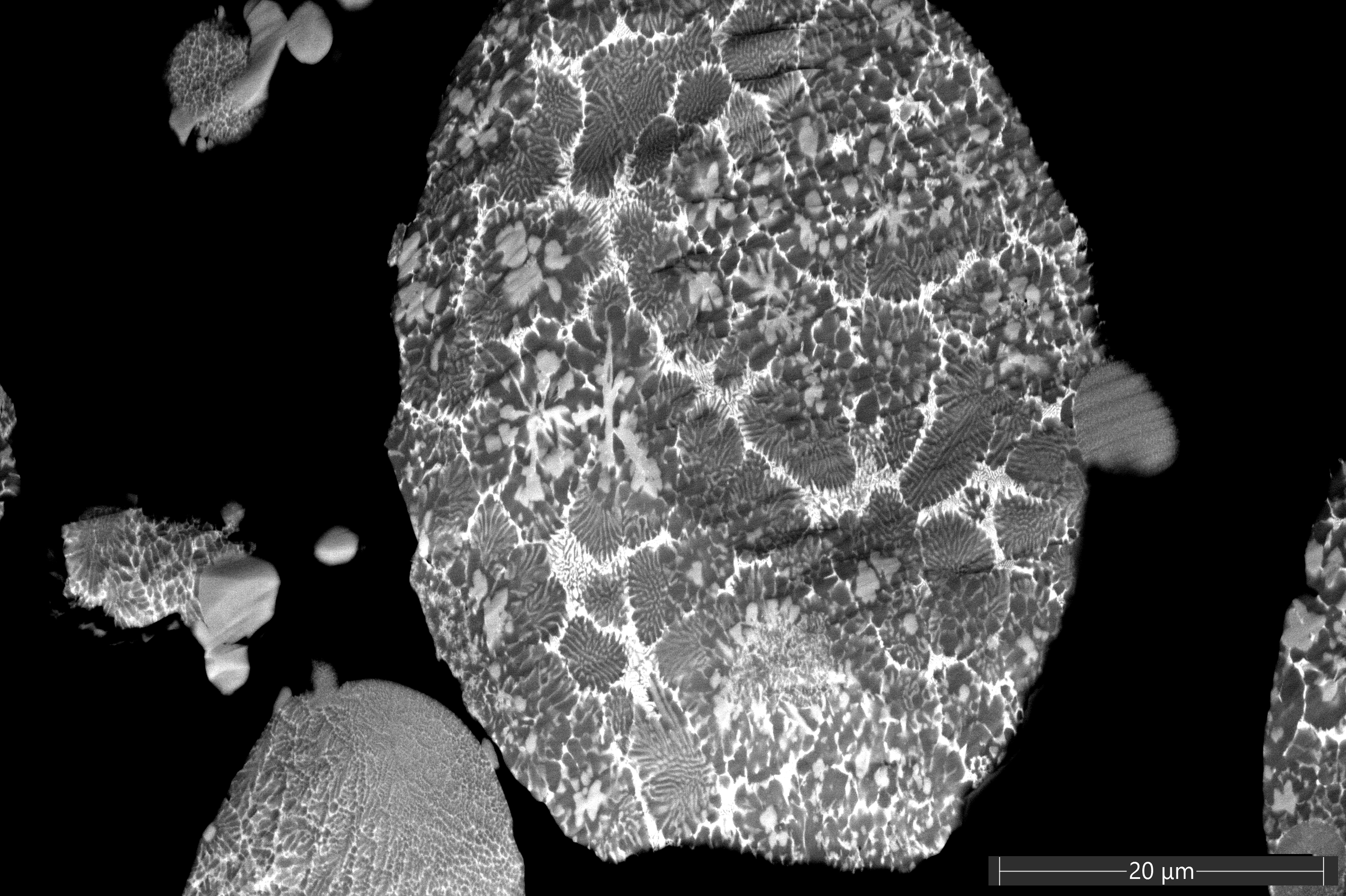
Alloy Garden
Artists: Anugrahaprada Mukundan (PhD student) and Mitsuhiro Murayama (Professor), Materials Science and Engineering Department, Virginia Tech
NNCI Site: NanoEarth
Tool: ThermoScientific Helios 5 UC Dual Beam
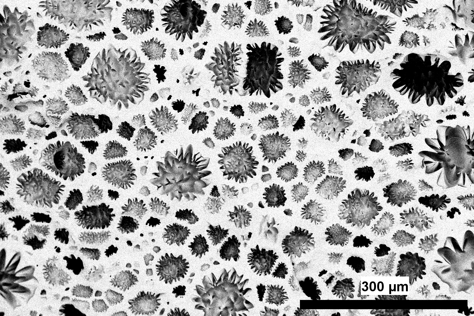
This scanning electron microscopy (SEM) image shows AlFeCe alloy powder particles, which were later used to 3D print the alloy. The aim of analyzing these powders is to explore potential correlations between the phases observed in the powders and those in the 3D-printed specimen. Within a single particle, the various phases appear in different sizes and morphologies, resembling flowers and trees in a garden, at the microscale.
NanoHearts: Self-Assembled Beauty at the Nanoscale
Artists: Sourav Sutradhar and Oghenetega Allen Obewhere (Grad students, Chemical and Biomolecular Engineering ,PhD Program.), University of Nebraska-Lincoln
NNCI Site: NNF
Tool: Scanning Probe Microscopy-Atomic Force Microscope (SPM-AFM)
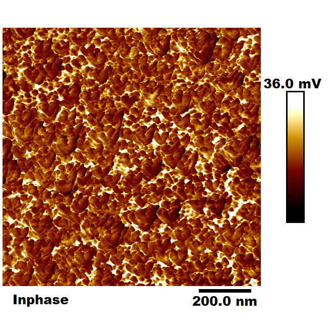
This AFM image reveals heart-shaped and granular surface features formed by a self-assembled monolayer of cystamine on a gold substrate. Captured at the nanometer scale, the image highlights densely packed, granular-shaped nanodomains with sharp phase contrast, indicating heterogeneous interactions across the surface. These nanoscale structures showcase the surprising elegance that emerges from molecular self-organization in engineered materials.
Foam in Bloom
Artists: Santhosh Sridhar (Ph.D. Student, Microcellular Lab Plastics Lab, UW ME), Ankush Nandi (Ph.D. Student, Vashisth Research Lab, UW ME), and Shaunak Deshpande (MS Student, Meza Research Lab, UW ME)
NNCI Site: NNI
Tool: ThermoFisher Scientific Apreo-S SEM
This striking micrograph captures the unique microstructure of vitrimer-based foams, resembling a landscape of blooming flowers frozen in time. Unlike conventional thermosets, vitrimers possess dynamic covalent bonds that enable reshaping, healing, and recycling without compromising integrity. When processed into foams, these adaptive networks form highly textured microstructures, with each spike and blossom reflecting the foam’s unique characteristics. The image highlights the unique ability of vitrimers to merge beauty with function, offering lightweight, recyclable materials that could transform structural applications while captivating the eye with their unexpected artistry.
Gold Herringbone
Artist: Zachery Enderson, Research Scientist II, Georgia Institute of Technology
NNCI Site: SENIC
Tool: Createc LT-SPM
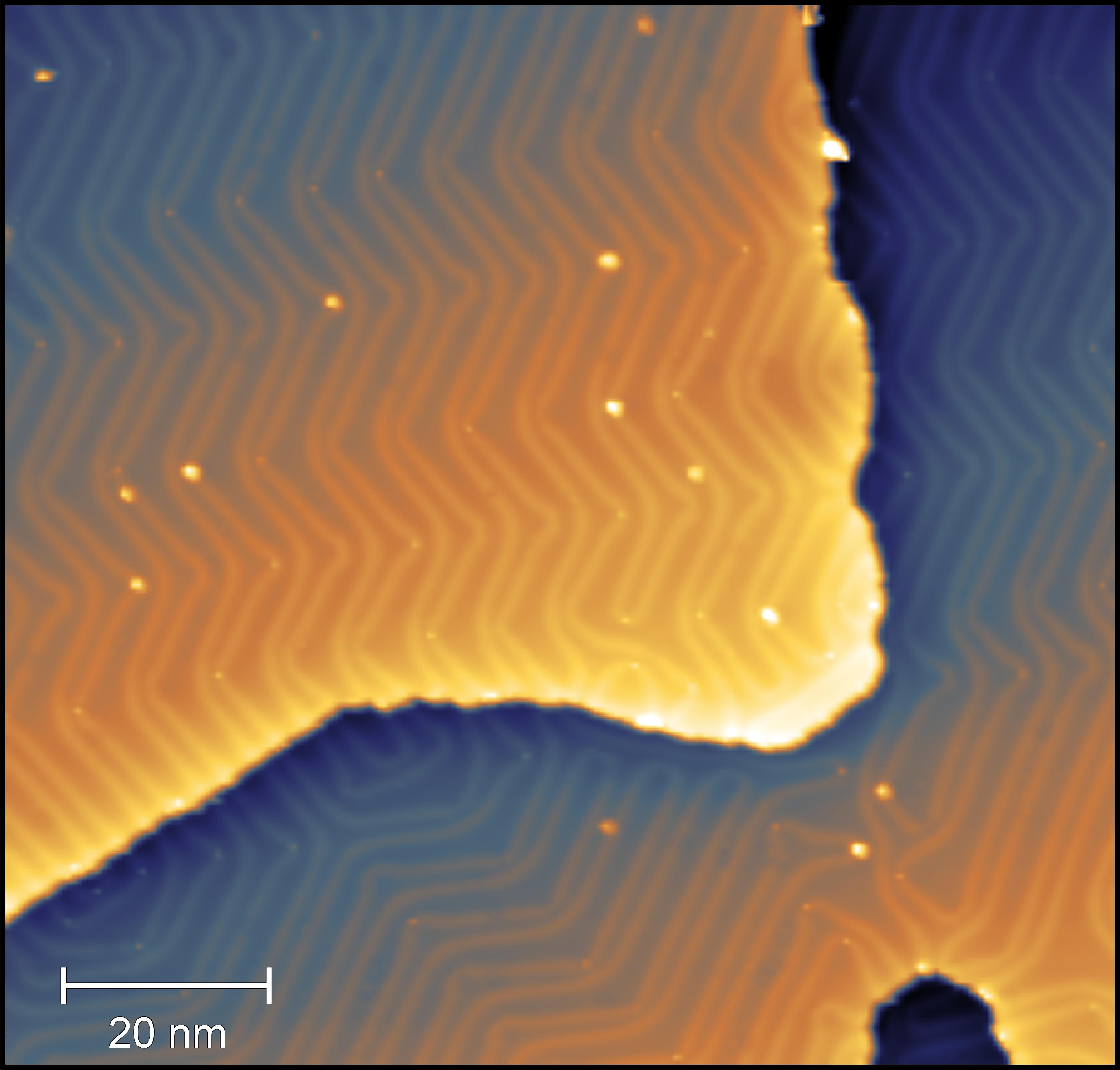
This scanning tunneling microscopy image shows the classic herringbone reconstruction of the Au(111) surface, recorded at liquid-helium temperature. The gold film, grown on mica and cleaned in ultrahigh vacuum immediately before imaging, exhibits a striking zig-zag pattern. This reconstruction arises from a slight mismatch between how the topmost layer of gold atoms prefers to arrange itself and the spacing enforced by the underlying layers. To relieve the resulting strain, the surface buckles into the herringbone motif, which can be seen continuing seamlessly across atomic step edges.
Incandescent Desert
Artists: Jack Hegarty, Graduate student, Vinayak Dravid Lab, Northwestern University
NNCI Site: SHyNE
Tool: Hitachi SU8700 SEM
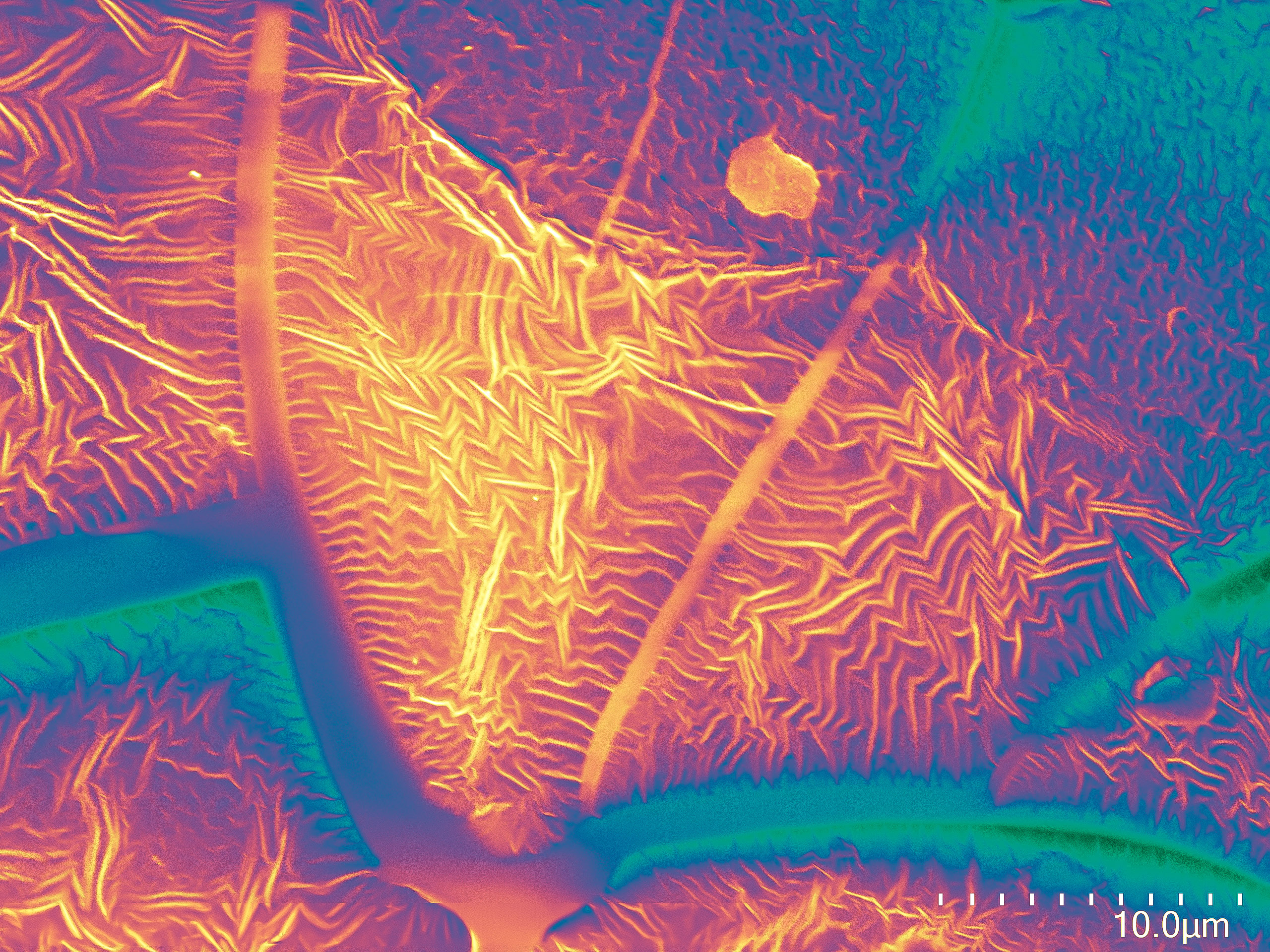
Scanning electron micrograph of an ion-exchange resin showing a wrinkled terrain of ~100-nm ridges. These materials are designed to efficiently capture atmospheric carbon dioxide when dry and release it when wet.