NNCI Image Contest 2024 - Unique
Most Unique Capability
This category celebrates the unique capabilities each site of the NNCI has on offer. Please check out the images below and read a little about the research behind them.
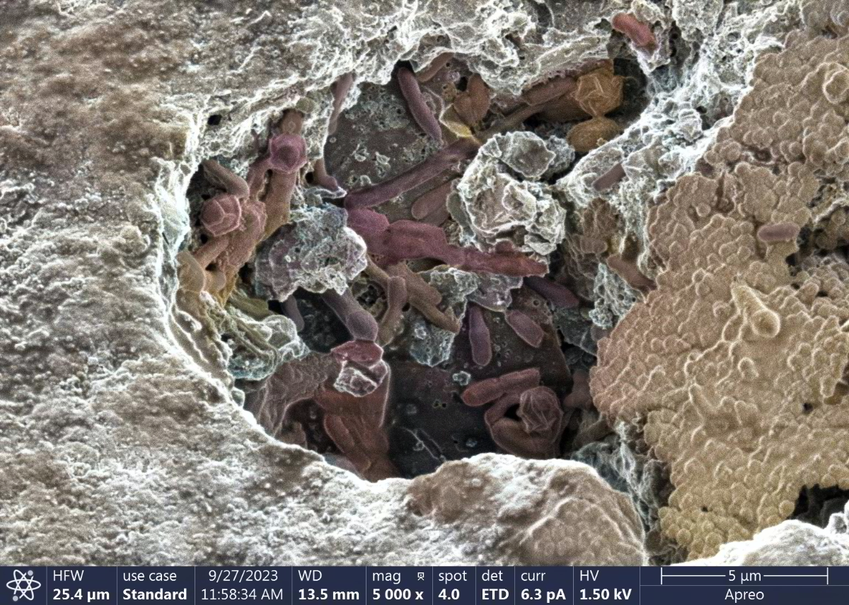
Microbes Hanging Out
Artist(s): Ed Winner (Vice President, Remediation Products, INC.), Jas Beharic & Julia Aebersold (UofL-MNTC Staff Members)
NNCI Site: KY Multiscale
Tool: SEM - Thermo Scientific Apreo
A microbial consortium growing within a surface fracture of granular activated carbon. This and similar pictures indicate that microbes, primarily bacteria, prefer to initiate growth in niches. The is colorized but otherwise unaltered.
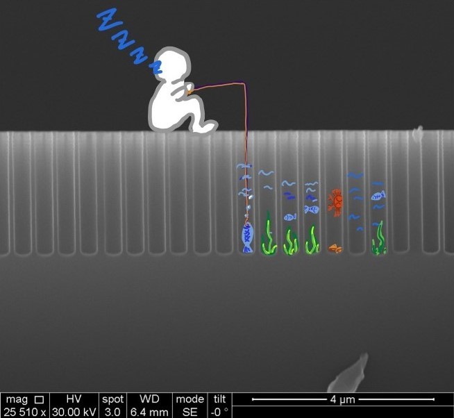
Fishing in the Wells
Artists: Tianyue “Mary” Gao & Langqiu Xiao, grad students, University of Pennsylvania
NNCI Site: MANTH
Tool: Quanta 600 FEG ESEM, SPTS Rapier Si DRIE, March Jupiter II Etcher
The image depicts a cross-sectional view of a silicon wafer after undergoing Bosch process. The aspect ratio of the wells is tunable. This array enables precise control of the microenvironment for catalytic reactions within the structures, allowing for the fine-tuning of reaction products in electrochemical processes.
Initially, a monolayer of polystyrene particles was assembled on the silicon surface. Following an oxygen plasma etch, the size of the particles was reduced, creating gaps between them. An aluminum oxide layer was then deposited over the particle array. After the particles were removed, deep reactive ion etching was performed to form vertical trenches.
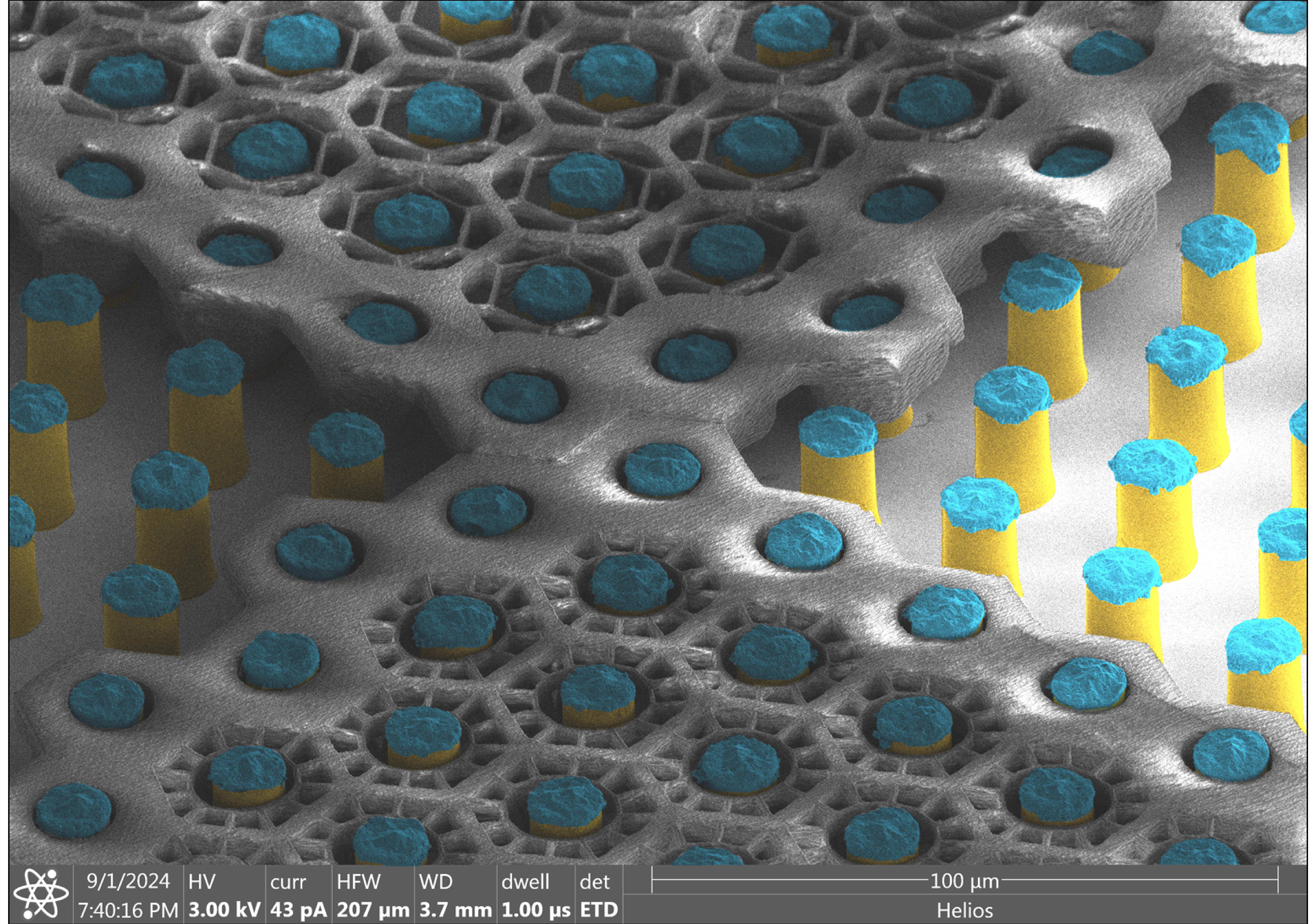
SubRetinal Prosthesis
Artists: Andrew Shin, PhD student, Stanford University
NNCI Site: nano@stanford
Tool: Nanoscribe GT, Heidelberg MLA150, EVG spraycoater, and Helios SEM
This SEM image shows a polymer scaffold built around pillar electrodes of the subretinal prosthesis designed for restoration of sight to the blind. A porous scaffold is intended to mimic a subretinal debris layer, which allows free flow of electric current, while preventing migration of the retinal cells. Electroplated gold pillars, shown in yellow, are 12 µm in diameter and 30 µm in height, coated with the sputtered iridium oxide, shown in blue. A polymer scaffold is printed around the pillars using 2-photon polymerization, featuring two designs - with 4 and 8 µm gaps.
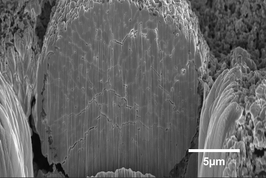
Advanced imaging of cathode active material degradation: insights through Helios 5 DualBeam system
Artists: Rupayan Ghosh (Graduate student) and Feng Lin (Associate Professor), Department of Chemistry, Virginia Tech
NNCI Site: NanoEarth
Tool: Helios 5 DualBeam System
The image, captured with the state-of-the-art Helios 5 DualBeam system, showcases the cathode active material after extensive charge-discharge cycling. In order to investigate mechanical degradation, we used precise focused ion beam (FIB) milling to reveal the internal structure of the material, exposing the cross-section of secondary particles. The image highlights crack formation, a critical insight into the material's failure mechanisms during cycling. Our group specializes in synthesizing and testing cathode materials under various conditions, followed by post-mortem analysis. The addition of the Helios 5 DualBeam system at NCFL has significantly enhanced our ability to investigate these phenomena, supporting the development of more robust materials and helping us validate our hypotheses.
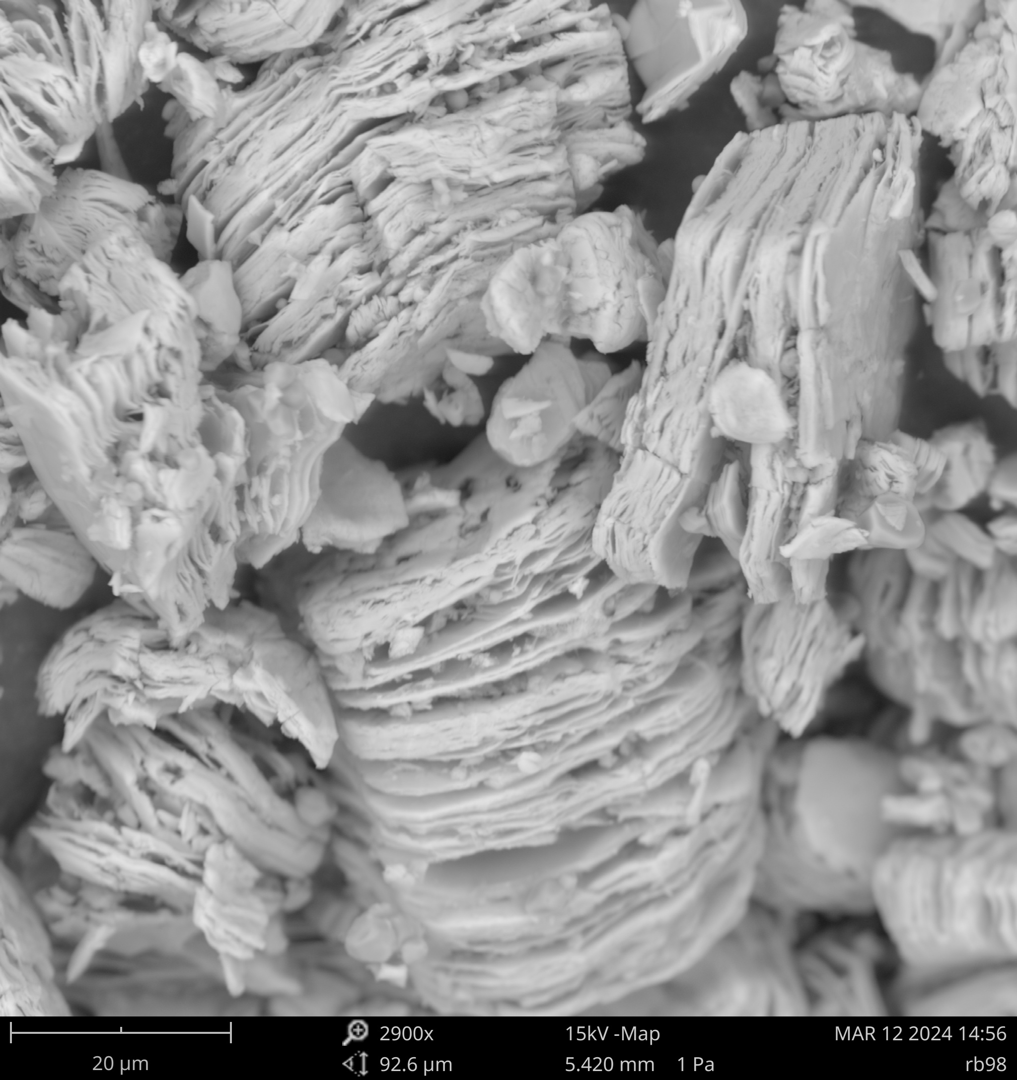
Vanadium Carbide (V2CTx) MXene
Artists: Reagan Beers, graduate student, Jessica Ray research group, Environmental Engineering, University of Washington
NNCI Site: NNI
Tool: Phenom ProX SEM
This image depicts a vanadium carbide (V2C) MXene made from acid etching of vanadium aluminum carbide. MXenes are two-dimensional nanomaterials with unique properties such as adjustable interlayer spacing and diverse chemical formulas. These properties are easily manipulated through chemical techniques and make MXenes prevalent in many applications including energy storage, photonics, and water treatment. In our lab, we have observed different structures and aqueous stabilities of the V2C MXene depending on the chemicals used in the synthesis. These differences will better guide other researchers to optimize their MXene for their desired application.
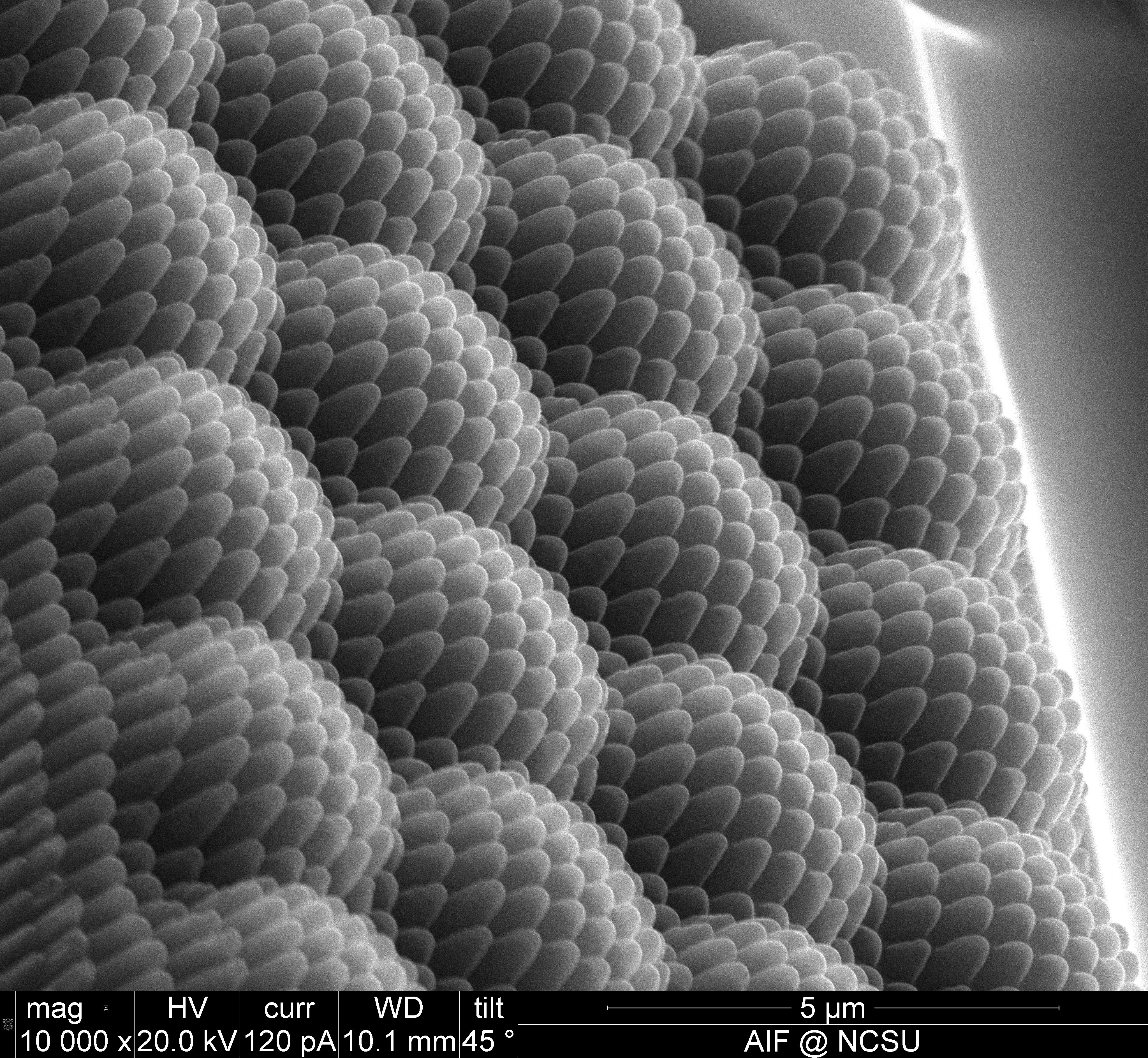
Hierarchical Diamond Die
Artists: Lauren Micklow, Senior Mechanical Engineer, Smart Material Solutions
NNCI Site: RTNN
Tool: FEI Quanta 3D DualBeam SEM/FIB
A diamond that has been patterned with a focused ion beam to create a dual scale texture. Following this small scale patterning process, the diamond with then be used as a stamp to replicate the pattern millions of times.
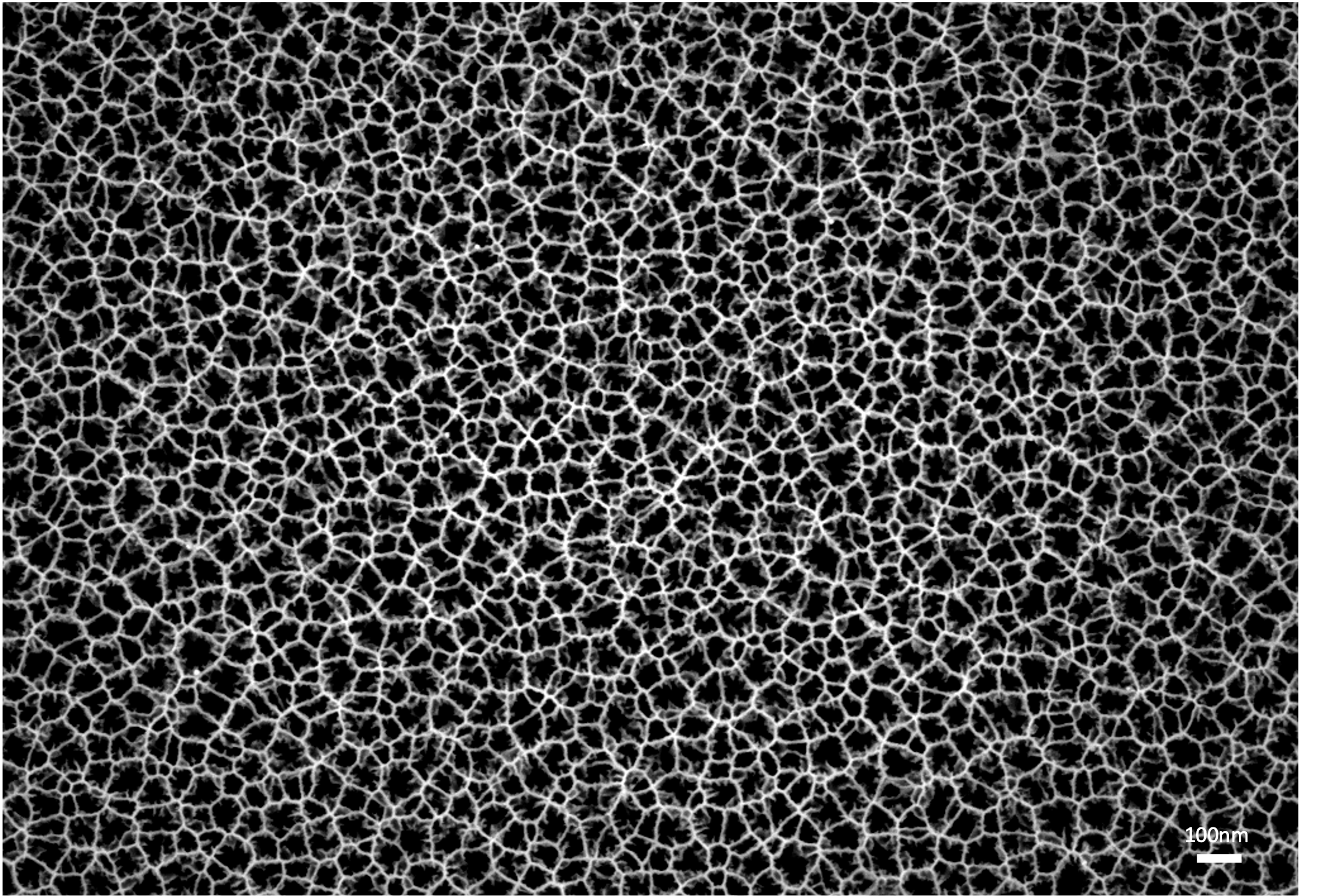
Detailed Pores
Artist: Gabriella Stark, Graduate Student, University of California - San Diego
NNCI Site: SDNI
Tool: Zeiss Sigma 500 SEM
This image depicts the top-down view of etched silicon. The etching process essentially electrochemically “drills holes” in the silicon to reveal each fine detail in the captivating porous structure shown.
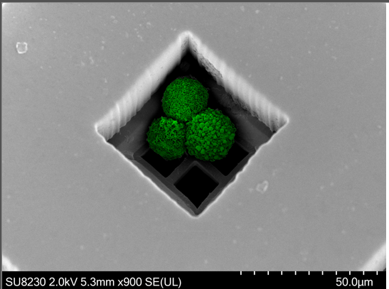
The Beauty of Cancer Cells
Artists Aref Valipour, Graduate Student, Sarioglu Group, Georgia Institute of Technology
NNCI Site: SENIC
Tool: Hitachi 8230
Image shows a cluster of cells captured with Cluster-Wells microdevices. We study the Circulating Cancer Cell Clusters (CTCCs) that are known to have more metastatic potential than single circulating tumor cells.
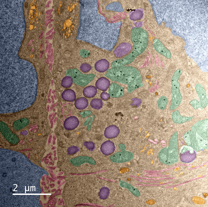
Cellowstone
Artists: Yi Li, Student, Kelley Research group, Northwestern University
NNCI Site: SHyNE
Tool: JEOL 3200
TEM image of a cell section. Nuclei: blue; filopodia: red; lysosomes: purple; mitochondria: green; cellular droplets: orange. The inner world of cells is as amazing as the outside world of Yellowstone National Park. Both are wonders created by nature. This work was carried out with the assistance of Dr. Reiner Bleher in TEM sample preparation, imaging, and naming.

