
NNCI Image Contest 2023 - Unique
Most Unique Capability
This category celebrates the unique capabilities each site of the NNCI has on offer. Please check out the images below and read a little about the research behind them.
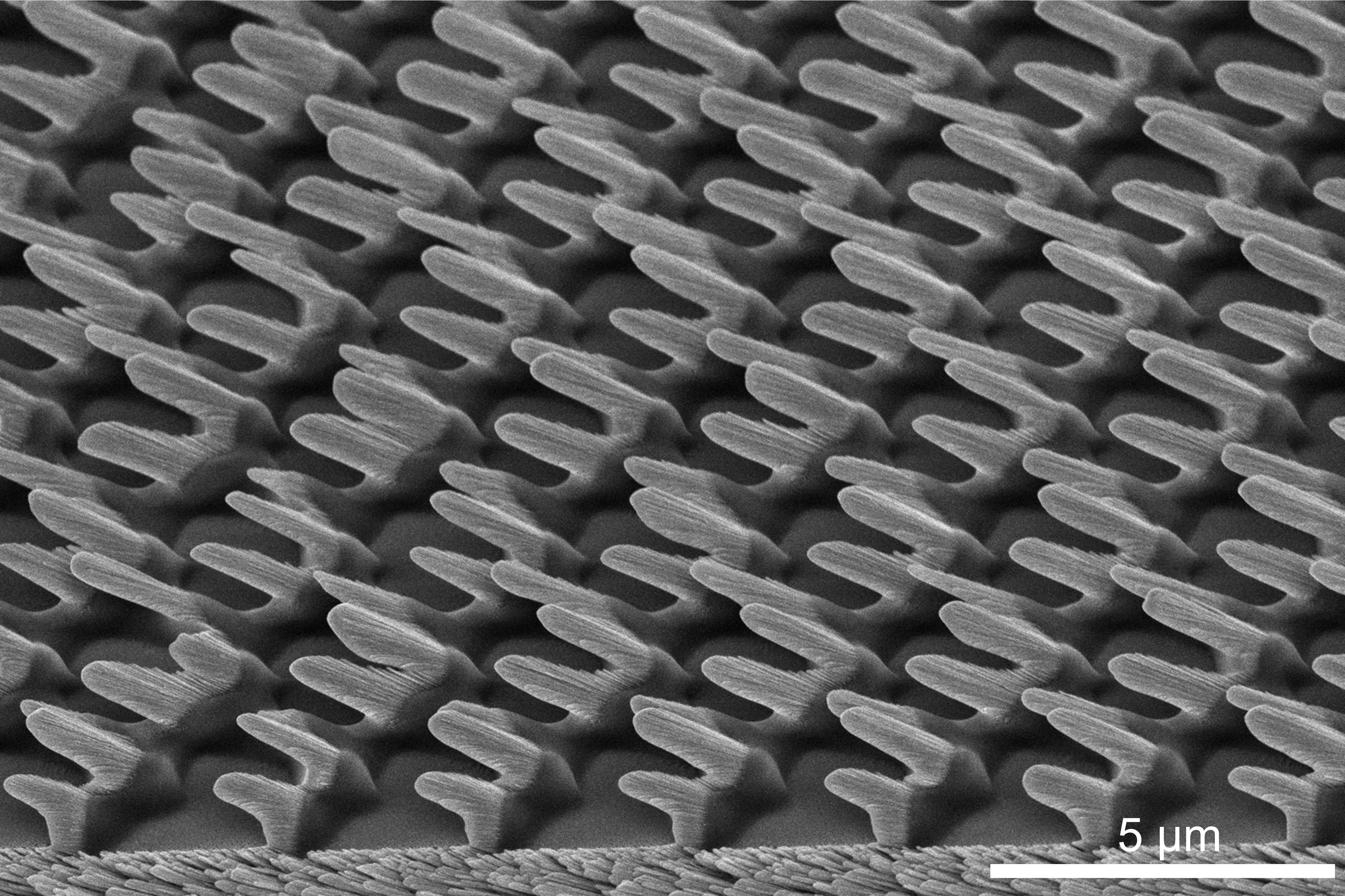
Shark Skin Mimicry
Artist: Chuang Qu, Research Scientist, University of Louisville
NNCI Site: KY Multiscale
Tool: Thermo Scientific Apreo SEM
This work presents the replication of shark skin dermal denticles using Glancing Angle Deposition (GLAD) atop line seeds. GLAD represents a highly advanced physical vapor deposition process that, when coupled with line seeds meticulously prepared through conventional photolithography, enables the creation of intricate three-dimensional micro/nanostructures on surfaces. These structures, in turn, impart exceptional properties, including superhydrophobicity, antifouling, and reduction in drag forces. The fabrication process was executed by Luca Caruso, a dedicated summer Research Experience for Undergraduates (REU) student, within the state-of-the-art cleanroom facilities at the University of Louisville.
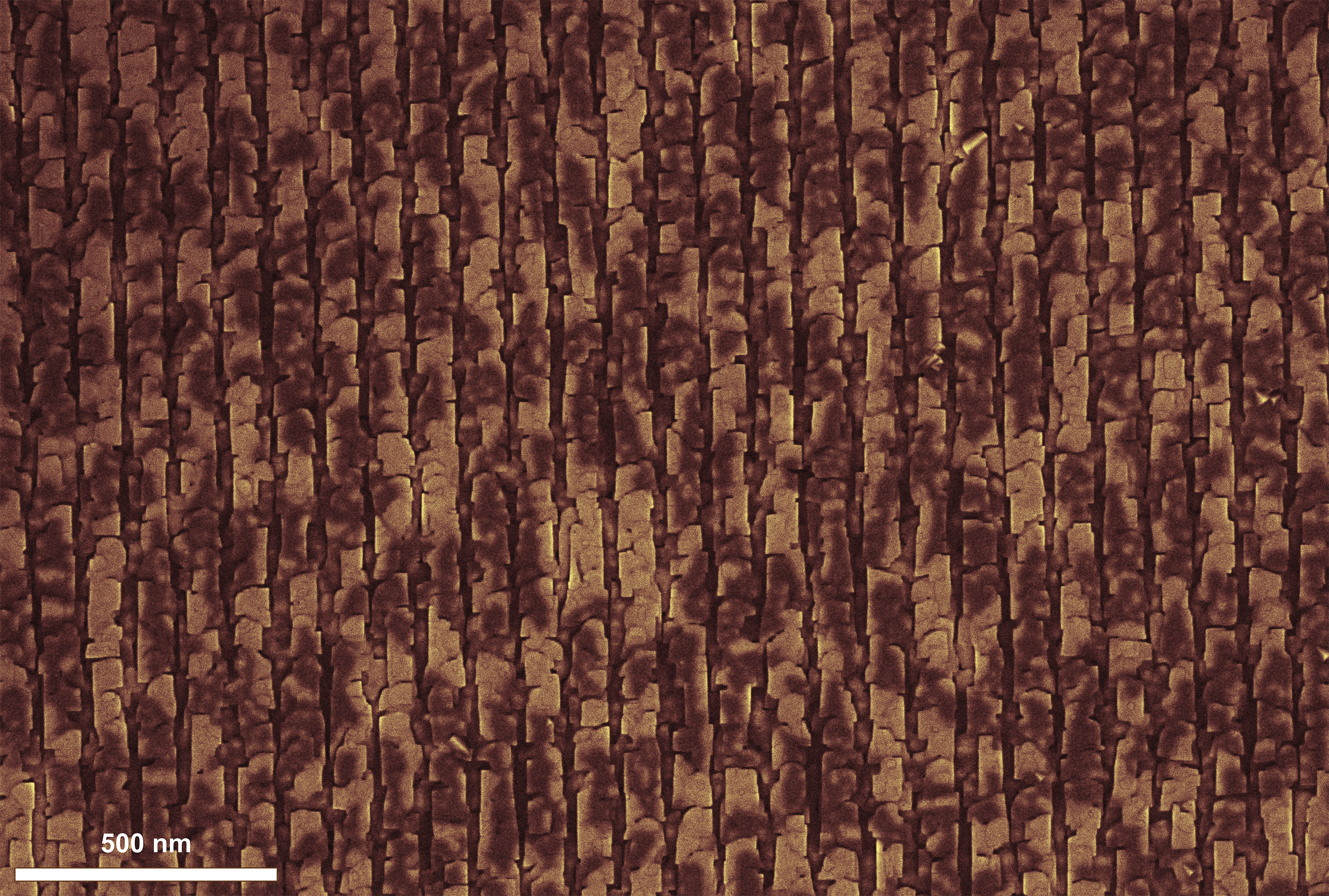
Conducting Nano Wooden Plank
Artist: Supriya Ghosh, Graduate Student, University of Minnesota
NNCI Site: MiNIC
Tool: Thermo Fisher Scientific Helios G4 UX Dual Beam FIB/SEM
The image shows the grains of a BaSnO3 film growing on patterned SrTiO3 substrate by hybrid molecular beam epitaxy. The substrate was patterned with vertical lines on the surface prior to film growth, by using a Ga ion beam to create channels in the material. As a result, when BaSnO3 is grown on top, the grains get aligned along this channel producing the “scale-like” effect. The channels are being used to study if we can assist the formation of “metallic” like defects in the host insulating thin film, to improve the conductivity of the thin film.
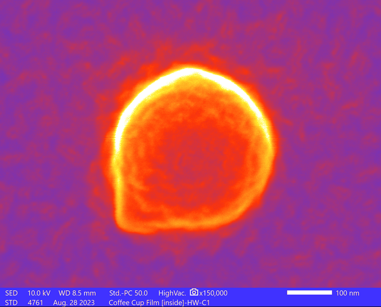
Nanoplastic Volcano Ball
Artists: Bipin D. Lade, (Postdoctoral Associate ) and F. Marc Michel, (NanoEarth Deputy Director, Associate Professor), Virginia Tech
NNCI Site: NanoEarth
Tool: JEOL IT-500HR analytical FEG SEM
The SEM image shows a single nanoplastic particle located on the surface of the inner lining of an unused single-use beverage cup. Micro-sized and nanosized plastic particles are released from these liners when exposed to water during use. Our laboratory is actively developing sampling protocols for nanoscale characterization and conducting research on nanoplastic synthesis and aggregation using hydrophobic fluids. Our primary focus lies in the characterization of nanoscale materials, and we utilize a range of analytical instruments, including transmission electron microscopy (TEM), scanning transmission electron microscopy (STEM), Raman spectroscopy, atomic force microscopy (AFM), dynamic light scattering (DLS), and UV-VIS spectroscopy.
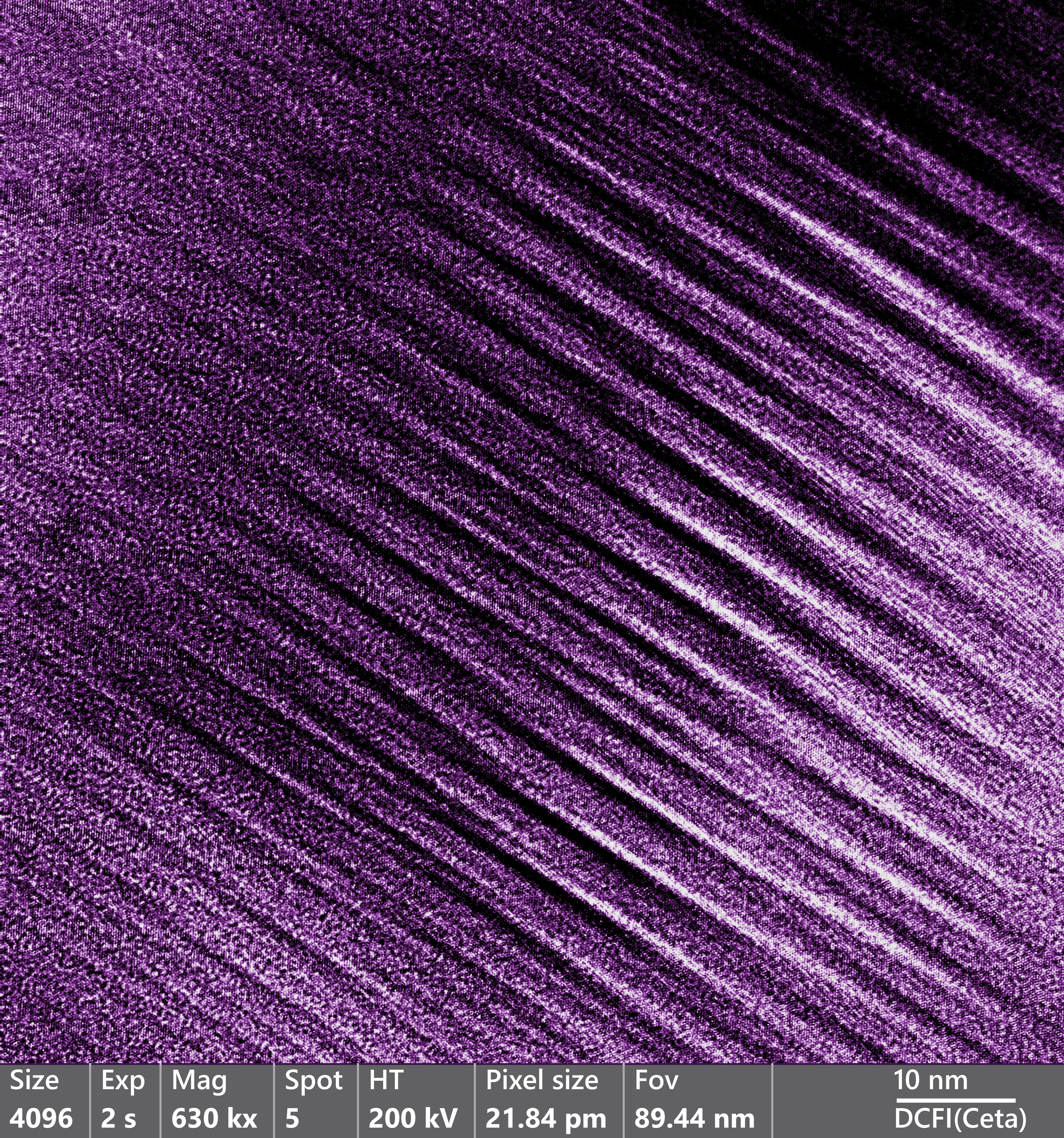
Atomic Structure of Carbon Chains at the Nanoscale
Artists: Darian Rosales (Undergraduate Student), Jesus Velazquez (Research Scientist), Blake Rogers (Graduate Student), Miguel José Yacaman (Professor), Northern Arizona University
NNCI SIte: NCI-SW
Tool: Transmission Electron Microscope
We worked this summer Northern Arizona University in the MIRA Center fabricating sp bonded carbon chains which are stabilized by gold atoms This image showcases the atomic structure of individual chains of carbon atoms. It was captured while conducting carbon nanomaterial attached to gold atoms. It was possible using the samples prepared during the summer to determine the way that the carbon chains crystallize. We combined HREM with Convergent beam diffraction to work out the structure.
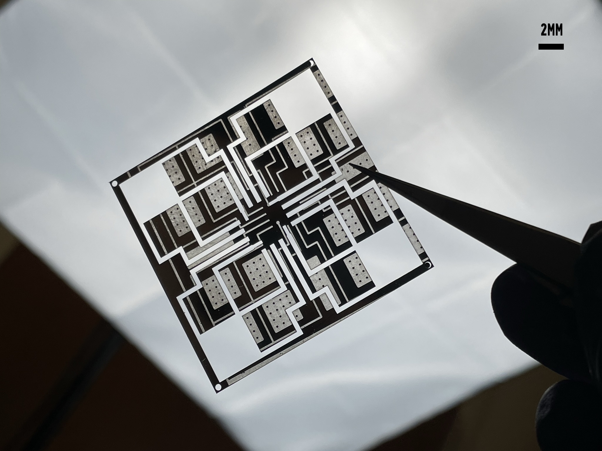
A Witness of Evolution
Artist: Kyle Pan, Graduate Student, Duke University
NNCI Site: Research Triangle Nanotechnology Network (RTNN)
Tool: CHA Industries Solution E-Beam Metal Evaporator & Suss MicroTec MJB3 Mask Aligner
This plastic sheet, with its futuristic look, is now a living canvas of our soft morphing robots' evolution. Its journey commenced as a mere glass cover sheet in the electron beam evaporation process, during the fabrication of our soft robot's sensing functionalities. As the design progressed, we embraced an air-pocket design, and this sheet transformed into a photolithography mask, resulting in its various patterns on the sheet. Now, as we further improved in the soft robot fabrication, this resilient sheet stands as a laser-cut mask for oxidized liquid metal, capturing the essence of progress in our research.
Influencing covalent network formation on surface using atomic hydrogen
Artists: Harshavardhan Murali, Graduate Student, School of Physics, Georgia Institute of Technology
NNCI Site: SENIC
Tool: Createc Low Temperature UHV Scanning Tunneling Microscope/Atomic Force Microscope
On-surface synthesis is a technique that can be used to grow clean two dimensional organic networks suitable for surface analysis in-situ at Ultra High Vacuum systems. Typically, the molecular deposition rate and substrate temperature are the only two parameters that can be adjusted during conventional growth. Here, we present a new method of adjusting the growth using atomic hydrogen generated in the chamber to inhibit the reaction, causing smaller organic networks (containing two and six membered units) to form and self-assemble. We demonstrate this effect using a triarylamine-based molecule as a model system. This technique may be used with other molecular systems as well, potentially unlocking a simple, yet powerful tool to control the synthesis.
Capturing Electrons
Artist: Serkan Butun, Research Associate, NUFAB, Northwestern University
NNCI Site: SHyNE
Tool: Nikon LV150
The image illustrates the trajectory of electron discharge within electron beam-sensitive resist on an insulating substrate. Notably, insulating substrates tend to accumulate surface electrons during electron probe processes like scanning electron microscopy and electron beam lithography, which unfortunately introduces artifacts like distortion, ultimately degrading the final output. In this particular instance, the surplus electron discharge takes center stage, elegantly surrounding a meta-lens structure. Meta-lenses, known for their capacity to focus light through engineered nanostructures, are showcased here. This remarkable work is done by I Tanriover and the Aydin Group on Raith Voyager100 at NUFAB, Northwestern University.
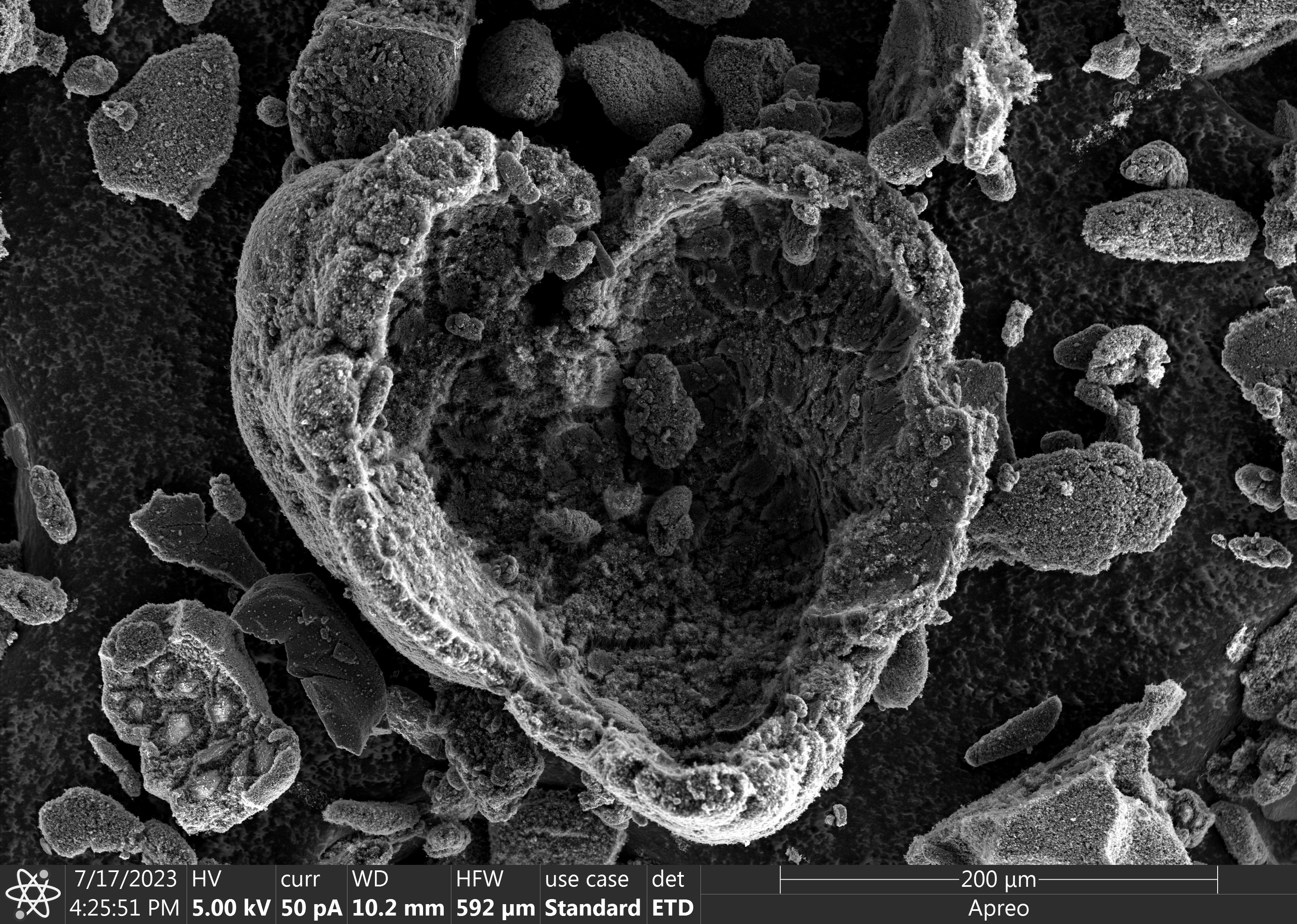
Carbon Heart
Artists: Henry Moise (grad student) and Marco Gigantino (post doc), Stanford
NNCI Site: nano@stanford
Tool: Thermo Fisher Scientific Apreo S LoVac SEM
Carbon nanotube shell grown around an iron catalyst bead, dislodged in the shape of a heart.