
NNCI Image Contest 2023 - Stunning
Most Stunning
This category celebrates the beauty of the micro and nanoscale. Please check out the images below and read a little about the research behind them.
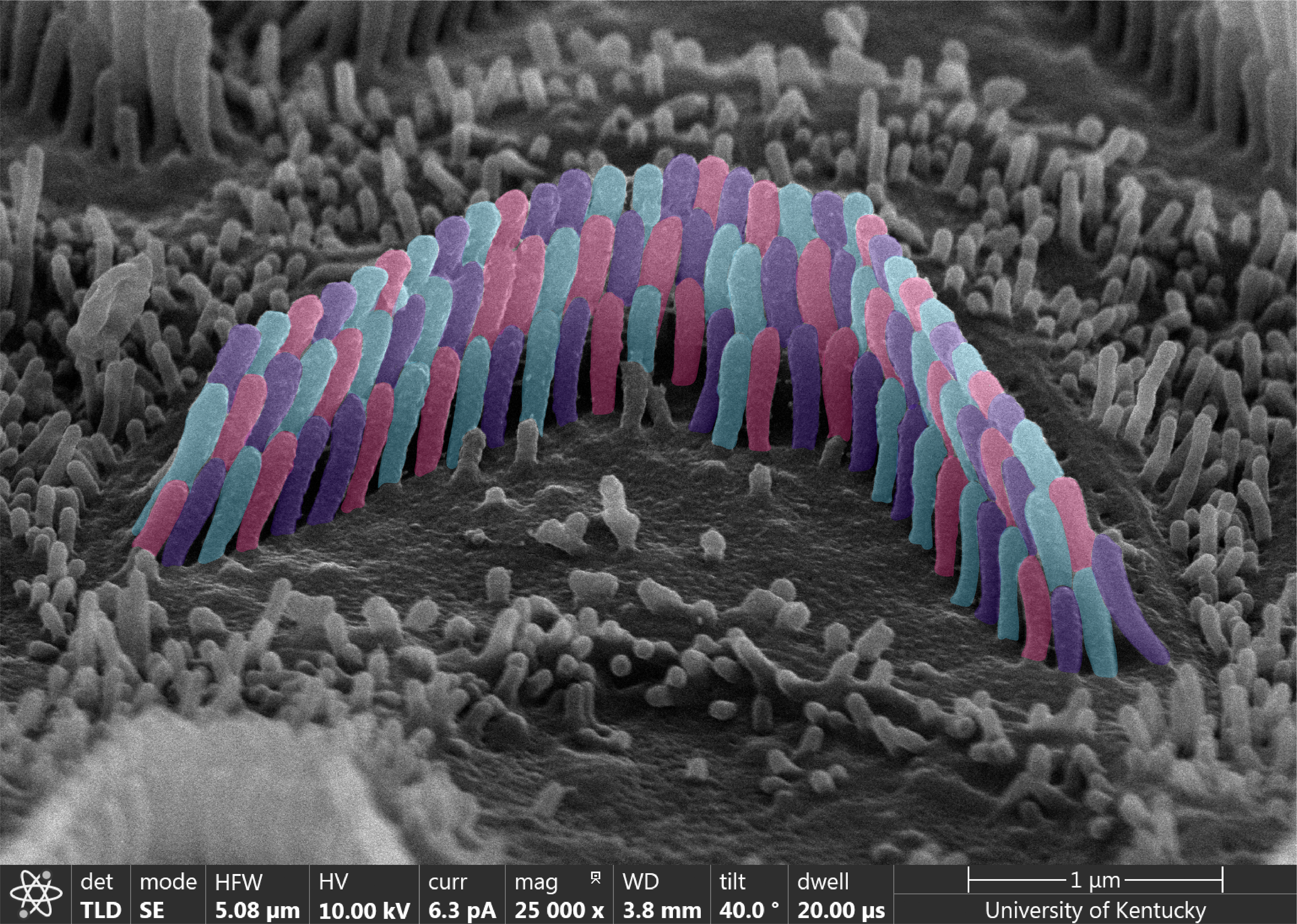
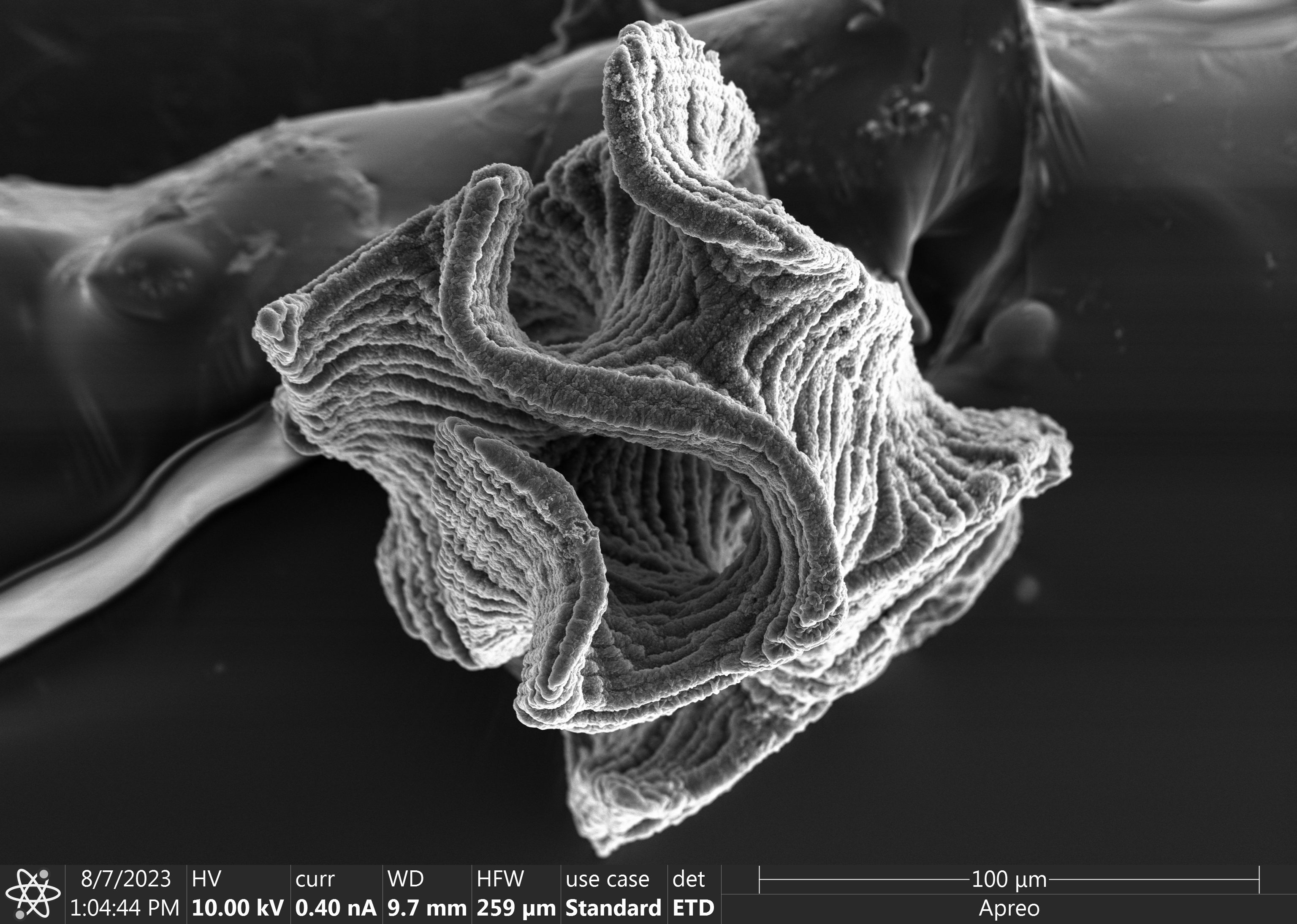
Colors of Sound
Artist: Abigail Dragich, Grad. Student, University of Kentucky
NNCI Site: KY Multiscale
Tool: Helios FIB-SEM
SEM image of an inner ear “hair cell” responsible for detecting sound within the cochlea. No one understands how these microstructures assemble and last over a lifetime of sound (since they cannot regenerate) making them highly researched in cell biology.
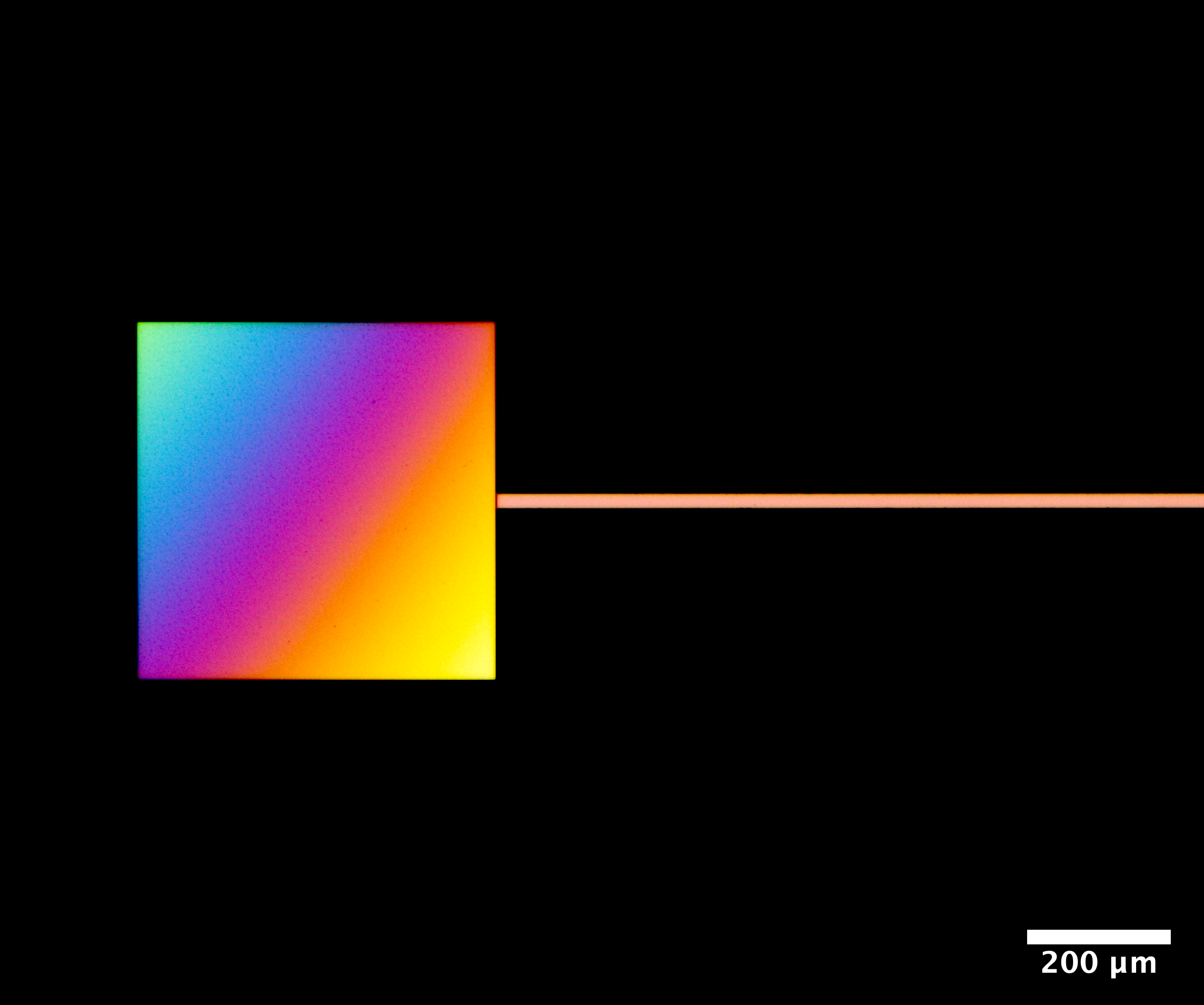
Balance
Artists: Matthew Campbell, Research Assistant Professor in MEAM, School of Engineering and Applied Science, University of Pennsylvania
NNCI Site: MANTH
Tool: Zeiss Axio Imager M2m optical microscope
The story behind this image is that at the edge of my wafer, my etch process didn't go completely through, and the thickness gradient of the remaining Silicon Dioxide film results in the coloration.
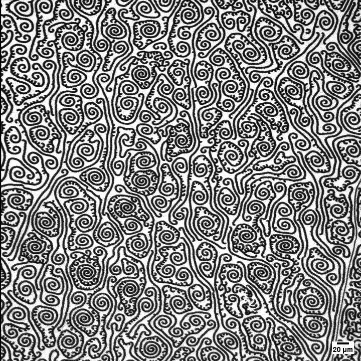
Lung Surfactant Swirls at the Bottom
Artist: Zach McAllister, graduate student, Chemical Engineering and Material Science, University of Minnesota
NNCI Site: MiNIC
Tool: Nikon Eclipse 80i
Every breath you take is possible because of lung surfactant. Lung surfactant has some very interesting properties and can rearrange itself to make these tiny, intricate, and beautiful patterns. The black patterns are more gel-like parts of the surfactant and the white background is the more liquid-like part of the surfactant. This image was made possible due to the gel-like part of the surfactant excluding a certain fluorescing molecule. Image taken by confocal fluorescence microscopy in the Zasadzinski Lab. Sample composition is 9:1 (4:1 r:racDPPC):hexadecanol with 1.5 mol% dehydrocholesterol.
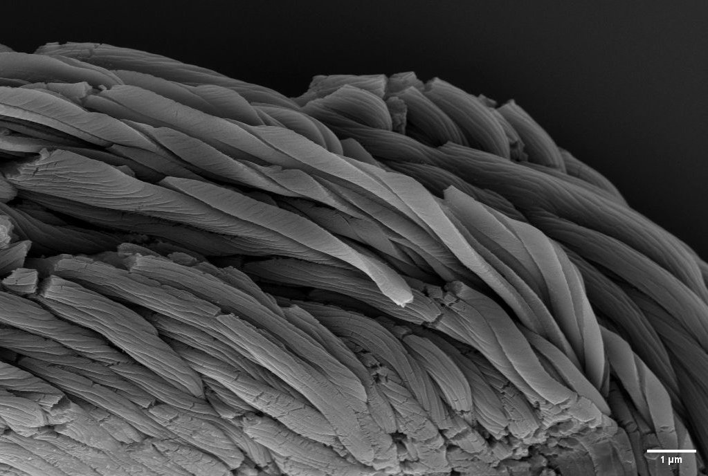
Micro-Resist Twist
Artist: Jay Graham, Undergraduate Research Assistant, Montana State University
NNCI Site: MONT
Tool: Zeiss SUPRA 55VP Field Emission Scanning Electron Microscope
This image is from a silicon sample that had been patterned with photoresist. The image shows an area of the sample where the photoresist had been disturbed, most likely during handling. The sample was used to evaluate the quality of the photolithography, but the photoresist structure seen in the image is not what is supposed to happen. The related research is in the development of fabrication processes to make improved nanostructured polarizers.
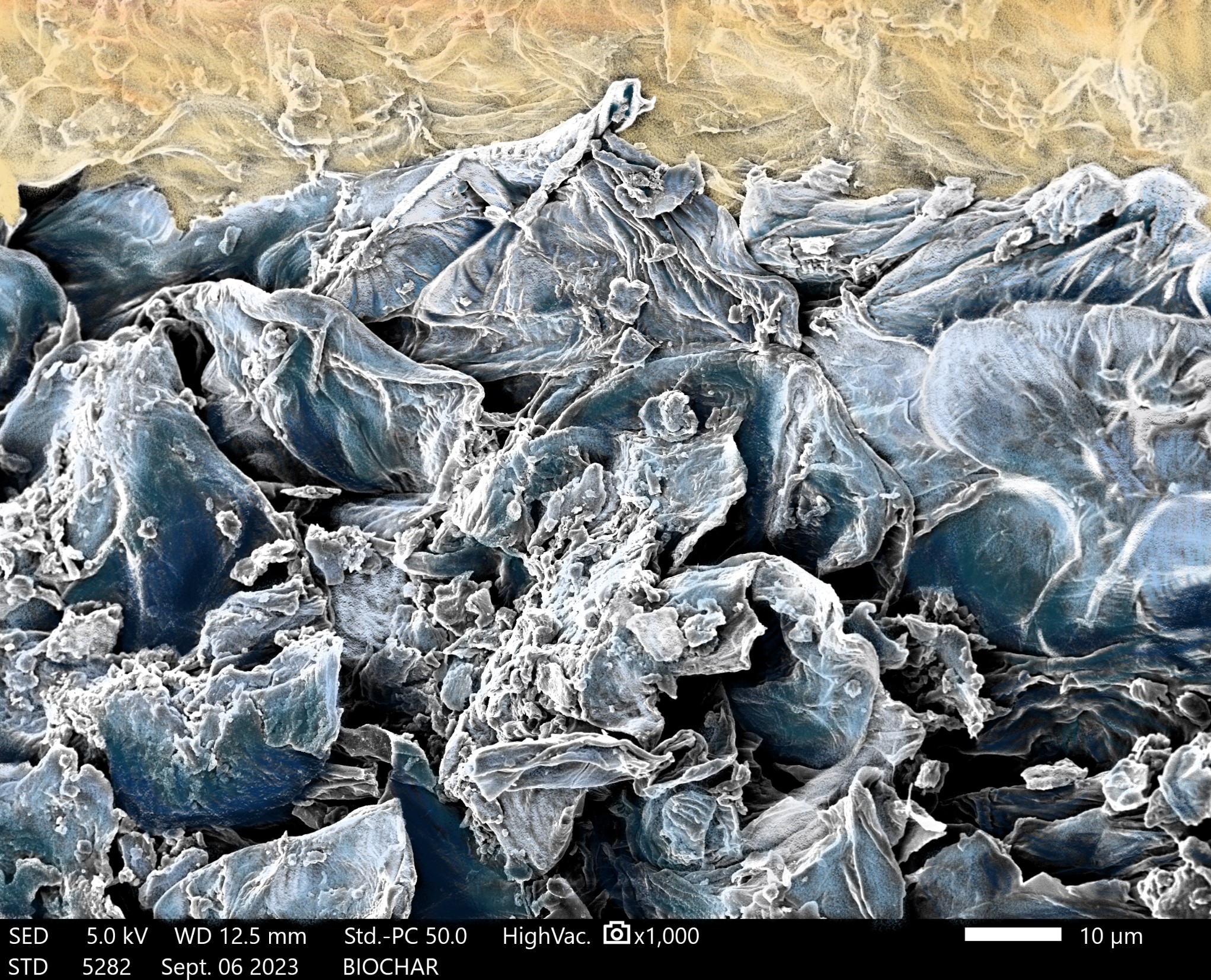
Surfing the Nano Seas
Artist: Yilin Li, Graduate Research Assistant, College of Agriculture Department of Food Science and Technology, Virginia Tech
NNCI Site: NanoEarth
Tool: JEOL IT500 SEM
The overall goal of the research is to develop a cost-effective filtration system that utilizes biochar produced from food waste to effectively reduce pathogens by adsorption. SEM images were used to assess the porosity and surface morphology of biochar derived from pecan shells carbonized at a temperature of 300 ºC. The images reveal a sheet-like structure, indicating potential for high surface area. SEM images enable the researchers to have a better understanding of the surface characteristics of biochar produced at a specific temperature and provide insight into its potential adsorption and filtration efficiency.
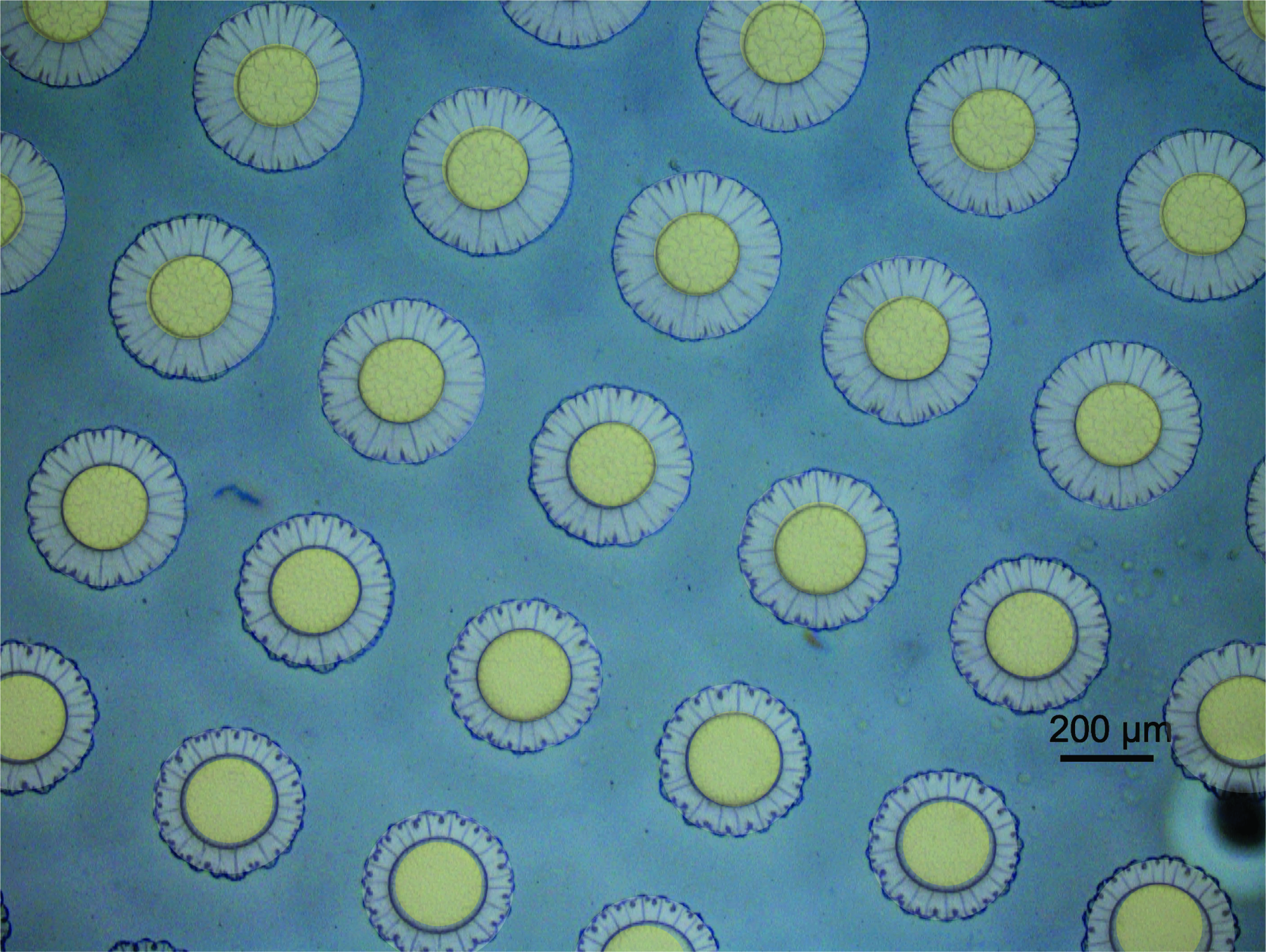
Blooming Hydrogels
Artists: Dr. Stephen Morin (Associate Professor), Nengjian Huang (graduate student), Brennan Watts (graduate student), Department of Chemistry, University of Nebraska-Lincoln
NNCI Site: NNF
Tool: Zeiss Axio Scope.A1
Micro-scale hydrogels attached to a support experience non-uniform strain during expansion. A mismatch between the relaxation rate of the polymer network and diffusion of water into the gel reveals flower-like structures. A visible diffusion front of water permeating the network gives the appearance of disk florets. This image was captured while researching the applications of micro-structured adaptive hydrogels bound to elastomeric supports.
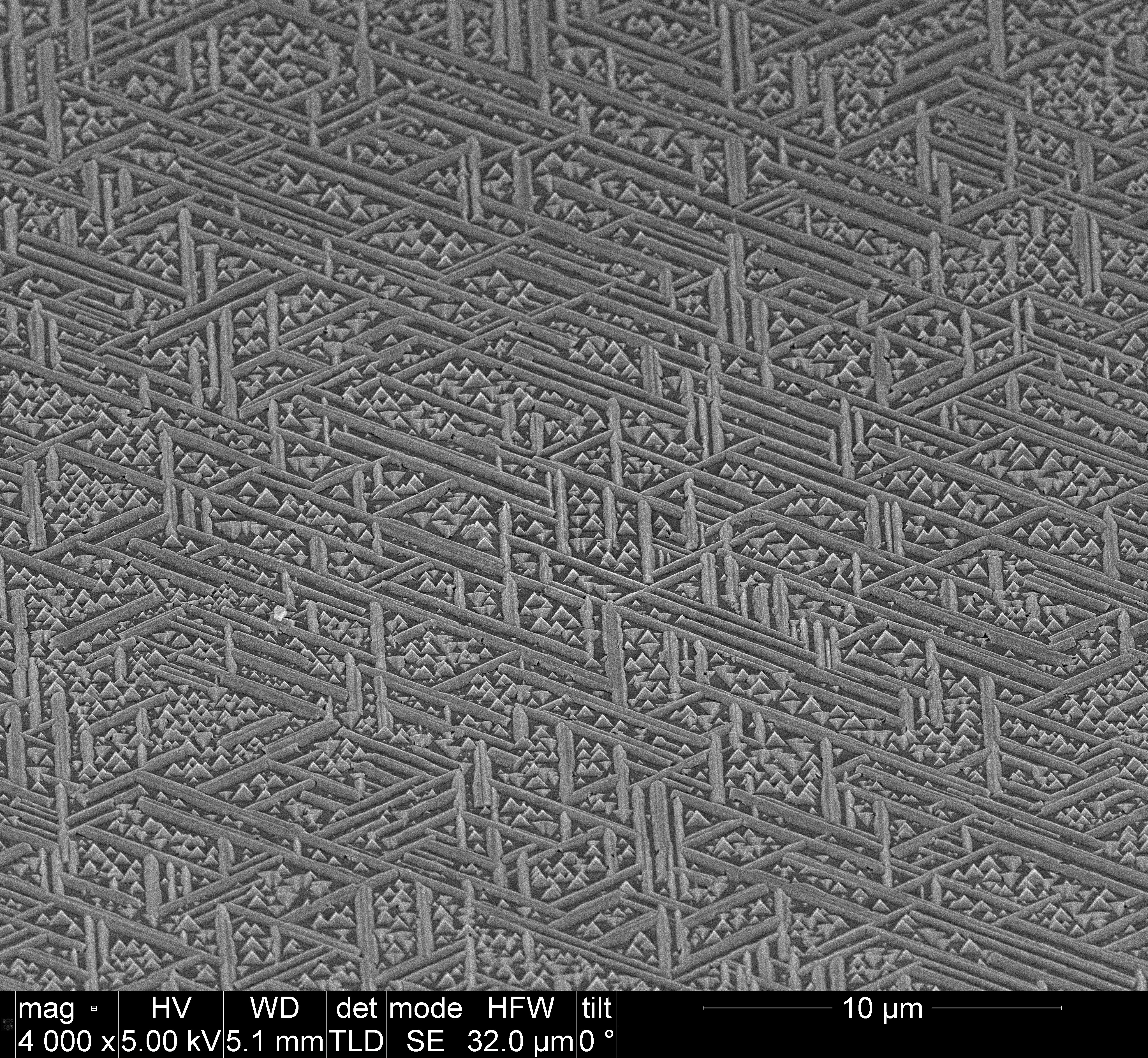
Perovskite Crop Circles
Artists: Alicia Bryan (Graduate Student - University of North Carolina Chapel-Hill), Aahan Dwivedi (Undergraduate Student - University of Florida)
NNCI Site: RTNN
Tool: FEI Helios 600 Nanolab Dual Beam System
Methylammonium lead iodide (MAPbI3) perovskite deposited on a mica substrate via chemical vapor deposition, demonstrating epitaxial rod-like structures and pyramidal crystallites. Image taken on FEI Helios 600 Nanolab Dual Beam System with a surface tilt of 45 degrees.
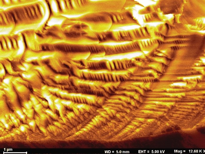
Stunning Wavy Resemblance
Artist(s): Sachin Shendokar (Grad student) and Shyam Aravamudhan, (Associate Professor), Joint School of Nanoscience and Nanoengineering
NNCI SIte: SENIC
Tool: Zeiss Auriga FIB/FESEM
NNCI Plenty of Beauty at the Bottom is an inspirational initiative always on my mind while investigating research responses. While investigating the Al2O3 degradation, the SEM image spontaneously felt like a cave surface. The stunning waves generated over the Al2O3 surface due to deionized water and silane flow were not only crisp in size but were also very ordered and gradual. The Luray Caverns resemblance incidentally has a similar acid water seepage theory behind the formation of the stalactites and stalagmites.

Candy Shop
Artist(s): Mike Barsoum, Graduate student, Northwestern University
NNCI Site: SHyNE
Tool: SEM FEI Quanta 650
This colorized scanning electron microscope image displays NU-1000, a unique metal-organic framework material. NU-1000, at the atomic scale, has a structure resembling a tiny, porous sponge, which can capture gases and find applications in gas storage and drug delivery with active Zirconium nodes. We are utilizing scanning electron microscopy to investigate its rod-like structure, uniformity, and presence surface defectiveness. This characterization was essential in determining the successful synthesis of this material and the particle size.
The Gyroidal Paradox
Artist: Jason Kronenfeld, grad student, Stanford
NNCI Site: nano@stanford
Tool: Thermo Fisher Scientific Apreo S LoVac Scanning Electron Microscope
A journey into the gyroid, a stunning shape plagued by the coexistence of simplicity and complexity. This geometry was fabricated while demonstrating our 3D printing research advancements in high-resolution continuous liquid interface production to produce complex particles en masse.