
NNCI Image Contest 2022 - Unique
Most Unique Capability
This category celebrates the unique capabilities each site of the NNCI has on offer. Please check out the images below and read a little about the research behind them.
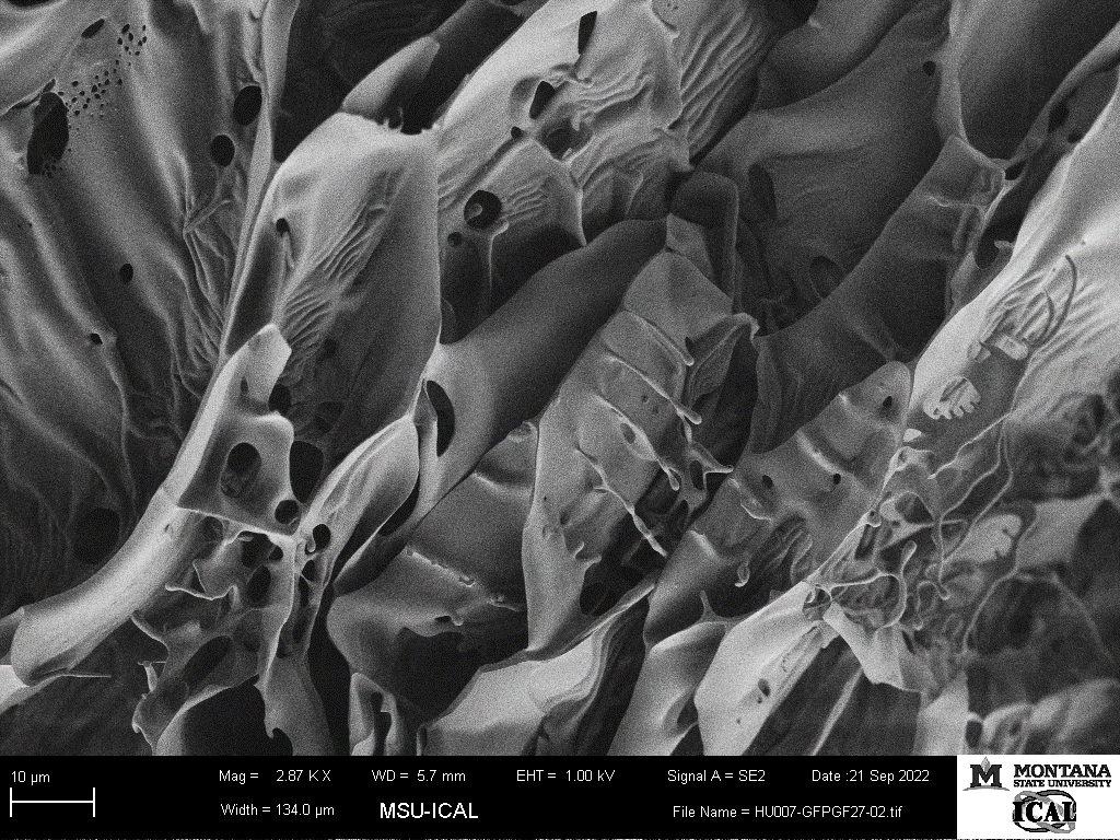
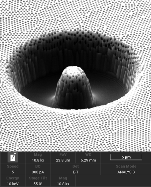
It’s slime! Cryo-SEM image of bioengineered human gastric mucus.
Artists: Katrina N. Lyon, PhD Student, Microbiology and Cell Biology, Montana State University; Bret Davis, Deputy Facility Manager ICAL, Montana State University
NNCI Site: MONT
Tool: Zeiss SUPRA 55VP Field Emission Scanning Electron Microscope using cryo-stage.
Bioengineered human gastric mucus grown in a stomach cell culture system for one month and imaged after flash-freezing in liquid nitrogen. The image shows the cleaved mucus surface, revealing the internal structure of the sample with its large honeycomb lattice and small mysterious drops on the edge of some of the lamellar structures. The cryo-stage of the FE-SEM allows for imaging of the sample with minimal drying or processing artefacts. This allows delicate structures such as the drops in this sample to remain for imaging.

Plenty of Waves at the Bottom
Artist: Mohammad Sayem Bin Abdullah, Graduate Student, University of Washington
NNCI Site: Northwest Nanotechnology Infrastructure (NNI)
Tool: SEM Apreo in the UW Molecular Analysis Facility
Large waves are hitting scattered mountainous islands during a tsunami. The waves are the gradual progression of a crack in additively manufactured (AM) Titanium. The tsunami is resembling the high-stress amplitude used during the fatigue experiment of the specimen. The crack grows at ~1 μm/cycle. This image confirms the ductile behavior in cyclic loading (fatigue), a prerequisite for large-scale manufacturing of titanium through AM processes. The specimen was built at 30 degrees using the Electron Beam Melting process, a novel AM technique to 3D print high-temperature metals. The geometric variability is being investigated to study the sustainability of the process.
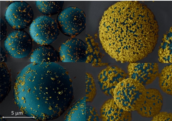
Detection of COVID-19 Antibodies
Artist: Neda Rafat, Postdoctoral Researcher, Parker H. Petit Institute for Bioengineering and Biosciences at Georgia Tech
NNCI Site: SENIC
Tool: SEM
Using polystyrene microbeads and a novel detection technique, COVID-19 positive serum samples (beads in the right column) were differentiated from healthy serum samples (beads in the left column).
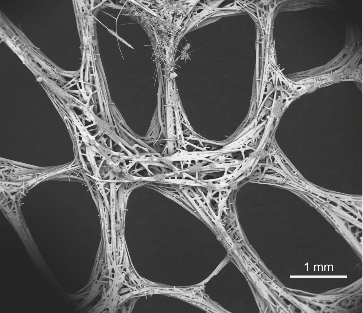
Biosilica fiber-based cellular structure of glass sponge’s sieve plate
Artist: Hongshun Chen, Ph.D. Candidate, Mechanical Engineering
NNCI Site: NanoEarth
Tool: FEI Quanta 600 FEG Environmental SEM
This backscatter image features the “weaving basket” made of biogenic silica, which is the sieve plate of the glass sponge Euplectella aspergillum. Its structure and mechanics can provide inspiration on the design of fiber-based cellular structure.
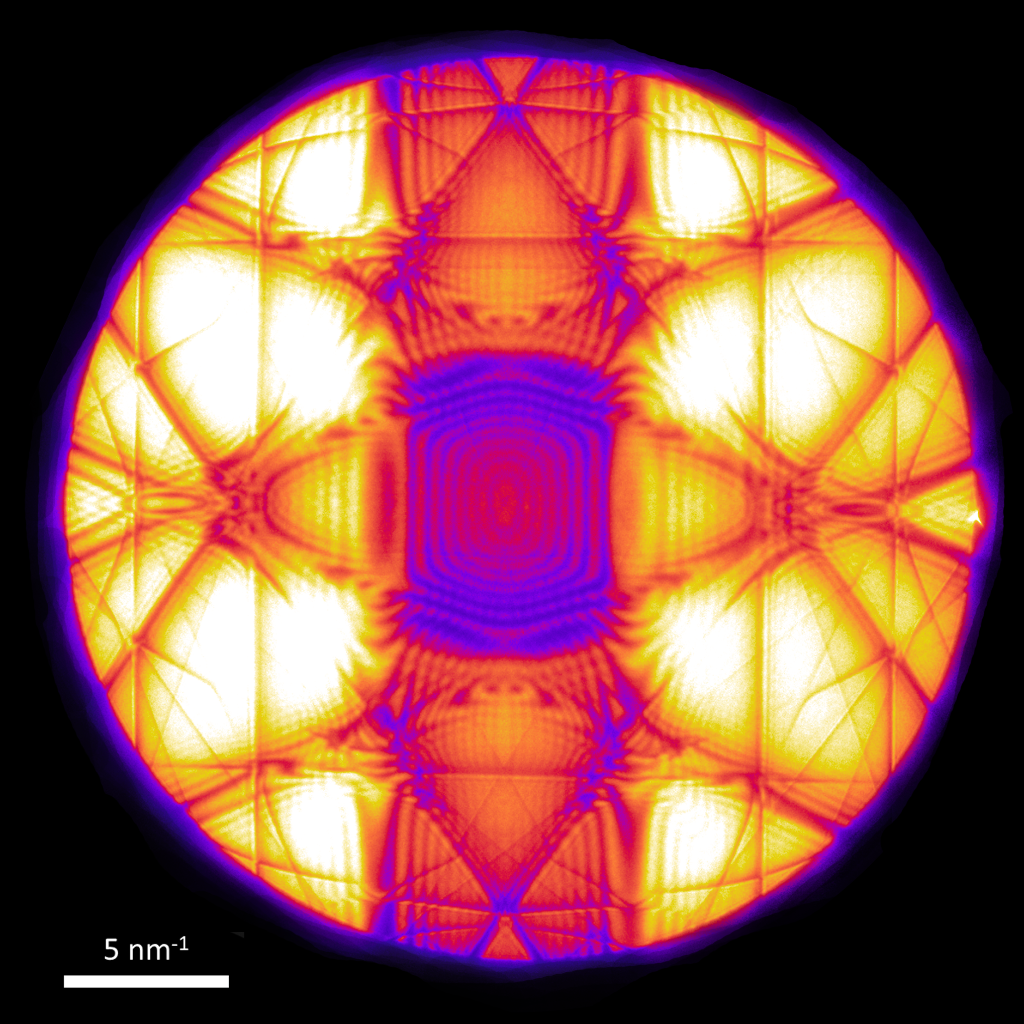
HypnoSis
Artist: Dr. Paul Smeets, Research Associate/TEM-FIB Facility Manager, NUANCE
NNCI Site: SHyNE
Tool: JEOL ARM300F (S)TEM
A standard piece of silicon (Si) can yield these beautiful and complex patterns with a converged electron probe. Specifically, large angle condensed beam electron diffraction (LACBED) makes it possible to obtain non-overlapping disks with larger diameter than the one determined by the Bragg angle. Beauty is in the eye of the beholder.. don't you Si?
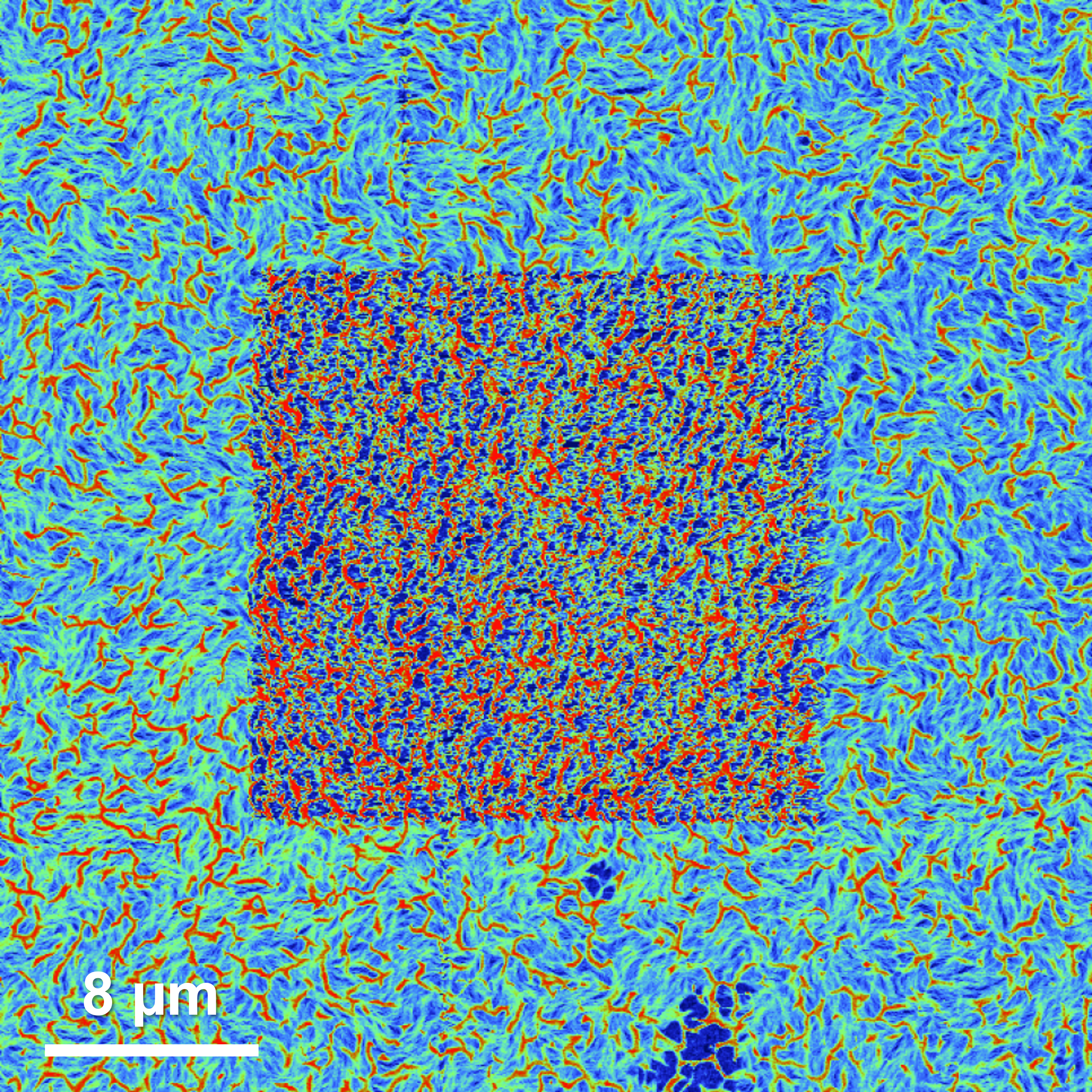
Below the Surface
Artists: Lukas Michalek, Post Doc and Kelly Lui, Graduate student, Stanford University-Zhenan Bao Group
NNCI Site: nano@stanford
Tool: Bruker Icon AFM
In the image the mechanical properties of a composite material consisting of a dielectric (dark blue regions) and a semiconducting polymer (red and green fibres) can be seen. The structure of the thin film was mapped with an atomic force microscope via quantitative nano-mechanical measurements. To be able to distinguish the structures below the surface a unique tip-based nanomachining approach was developed and executed in the centre of the image (square). A higher concentration of softer dielectric was recorded after removing the top layer with the same instrument. The findings help support the development of novel materials for flexible electronics.
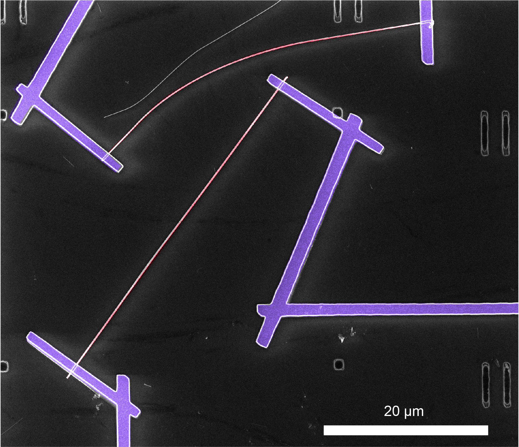
Sunlit Nanowires
Artist: Samuel Bottum, Graduate Student, UNC Chapel-Hill
NNCI Site: Research Triangle Nanotechnology Network (RTNN)
Tool: FEI Helios Nanolab 600, KJL PRO Line PVD 75 E-beam evaporator, Alcatel AMS 100, NPGS (software)
This image depicts a device fabricated to measure the photovoltaic properties of single multijunction silicon nanowires. This process involves making metal contacts (purple) to silicon nanowires (red) on a marker pattern (grey numbers), which are etched into the substrate. Each device holds ~20 nanowires with two contacts to each wire. The process to make this device involves many CHANL capabilities, including e-beam lithography, e-beam evaporation, DRIE, and SEM.
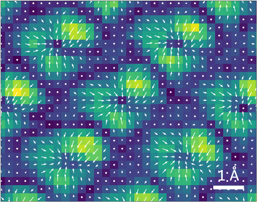
SrTiO3 4DSTEM Map
Artist: Jules Gardener, Senior Scientist, Harvard University & Austin Akey, Senior Scientist, Harvard University
NNCI Site: Center for Nanoscale Systems (CNS)
Tool: TEM-11 JEOL ARM
This image shows the electron beam deflection at ultra-high resolution of the SrTiO3 lattice. This deflection is due to the local electric field of the atomic columns and provides a direct method to visualize this field at the sub-atomic length scale. Arrows represent the direction and size of the displacement, while color represents displacement magnitude. The electron beam deflection was measured using a method called 4D scanning transmission electron microscopy (4D-STEM) center of mass (COM) in which a diffraction pattern is acquired and analyzed at each pixel.
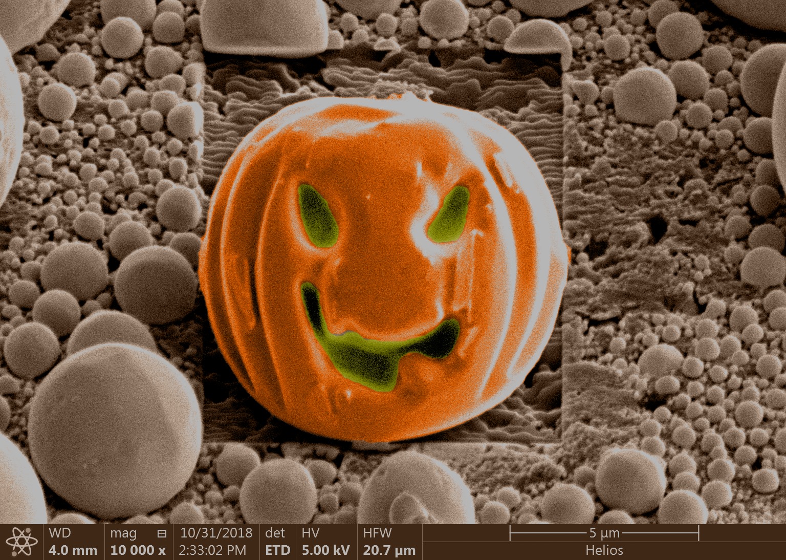
A Small Halloween Treat
Artist: Dr. Nicholas Seaton, Research Scientist, University of Minnesota
NNCI Site: MiNIC
Tool: Thermo Fisher Scientific Helios G4 UX Dual Beam FIB/SEM
This image was made shortly after the install of the Thermo Fisher Helios G4 UX FIB/SEM. I was practicing with some of the patterning features and realized I could turn one of the tin balls used for calibration of the beam into a very small pumpkin for Halloween. This demonstrates the excellent quality of the ion beam and the fine precision cuts it make. This is a colorized version of the original greyscale image.
Sombrero - Mexican Hat
Artist: Pariasadat Musavigharavi, Postdoctoral Fellow, University of Pennsylvania
NNCI Site: MANTH
Tool: TESCAN S8000X FIB/SEM
In order to improve the performance of the porous Silicon electrodes, Silver (Ag) nanoparticles are introduced to nanowells array of Silicon electrode. These Ag nanoparticles are blocked/covered by the walls of the arrays. FIB/SEM is a powerful technique to un-reveal the structure and chemical composition of nanoparticles by milling a few nanowells and capturing the inside.
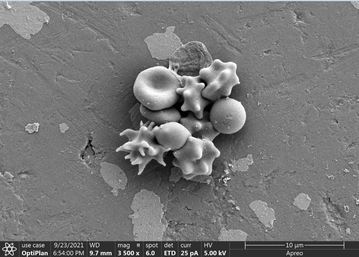
Fifty Shades of Red Blood Cells
Artist: Charles Elder, Grad Student, University of Louisville
NNCI Site: KY Multiscale
I am developing a method to freeze Red Blood Cells so they can be preserved for possibly decades. During freezing the cells are loaded with compounds that are foundin the freezing solution. We believe this does not happen in a uniform fashion with some cells being loaded withmore compounds than others. In this SEM image you can see that the cells are under different stresses which iswhy some look normal, and others look dead or extremely hypertonic. This may be because these cells wereloaded with different amounts of compounds from the freezing solution.
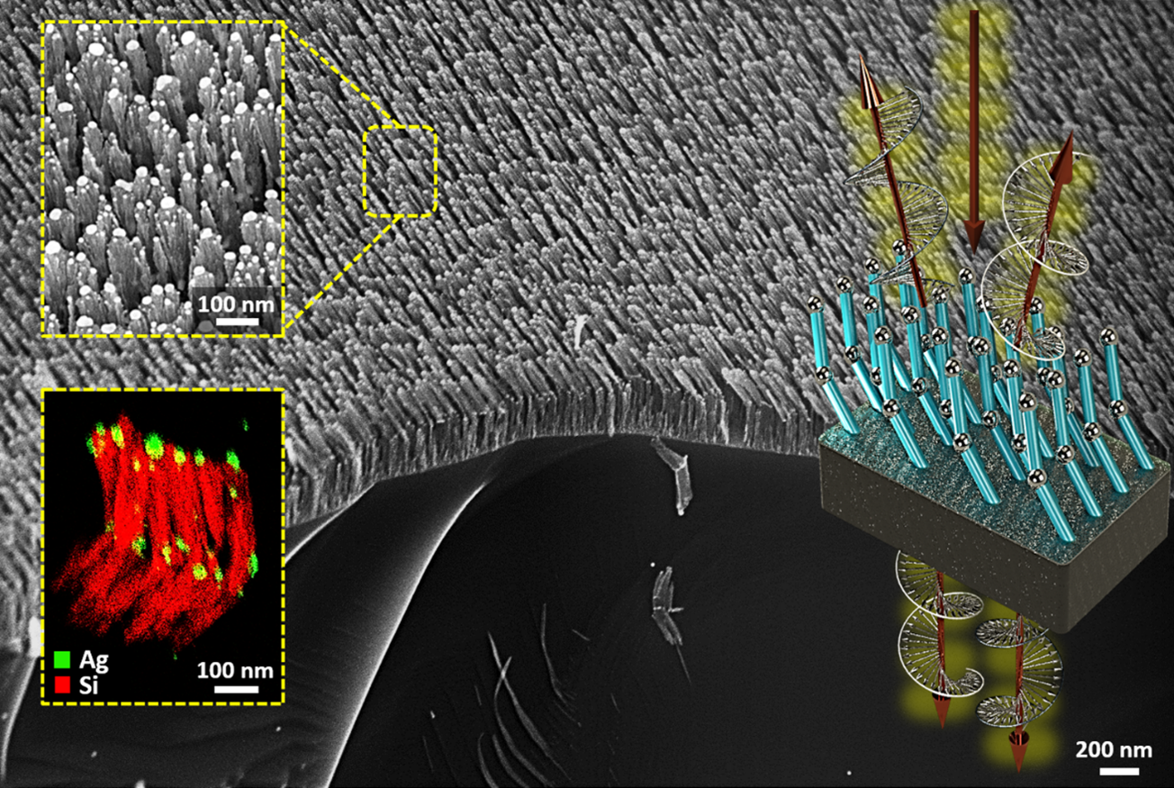
Nanoboomerang Forest
Artists: Shawn Wimer (Graduate Student), Dr. Ufuk Kilic, and Dr. Matthew Hilfiker, Electrical and Computer Engineering Department, University of Nebraska Lincoln
NNCI Site: Nebraska Nanoscale Facility (NNF)
Tools: FEI Helios NanoLab 660 and FEI Tecnai Osiris (S)TEM
Recently, there has been a paradigm shift in the engineering of optical materials from single material building blocks to the hybridized metamaterial platforms which can unlock new possibilities in the manipulation of chiral light-matter interactions at nano scale. This collage image shows our new metamaterial design: wafer-scale-area deposited, spatially coherent, super-lattice type, three-dimensional, distorted L-shape plasmonic hybrid metamaterial so-called plasmonic Nanoboomerang. Here, the scanning electron microscope image (SEM) from the tilted cross section of the sample was shown with the top view SEM image and the Scanning-Transmission-Electron-Microscopy image with atomic resolution energy-dispersive X-ray spectroscopy-based material mapping as insets.