
NNCI Image Contest 2022 - Stunning
Most Stunning
This category celebrates the beauty of the micro and nanoscale. Please check out the images below and read a little about the research behind them.
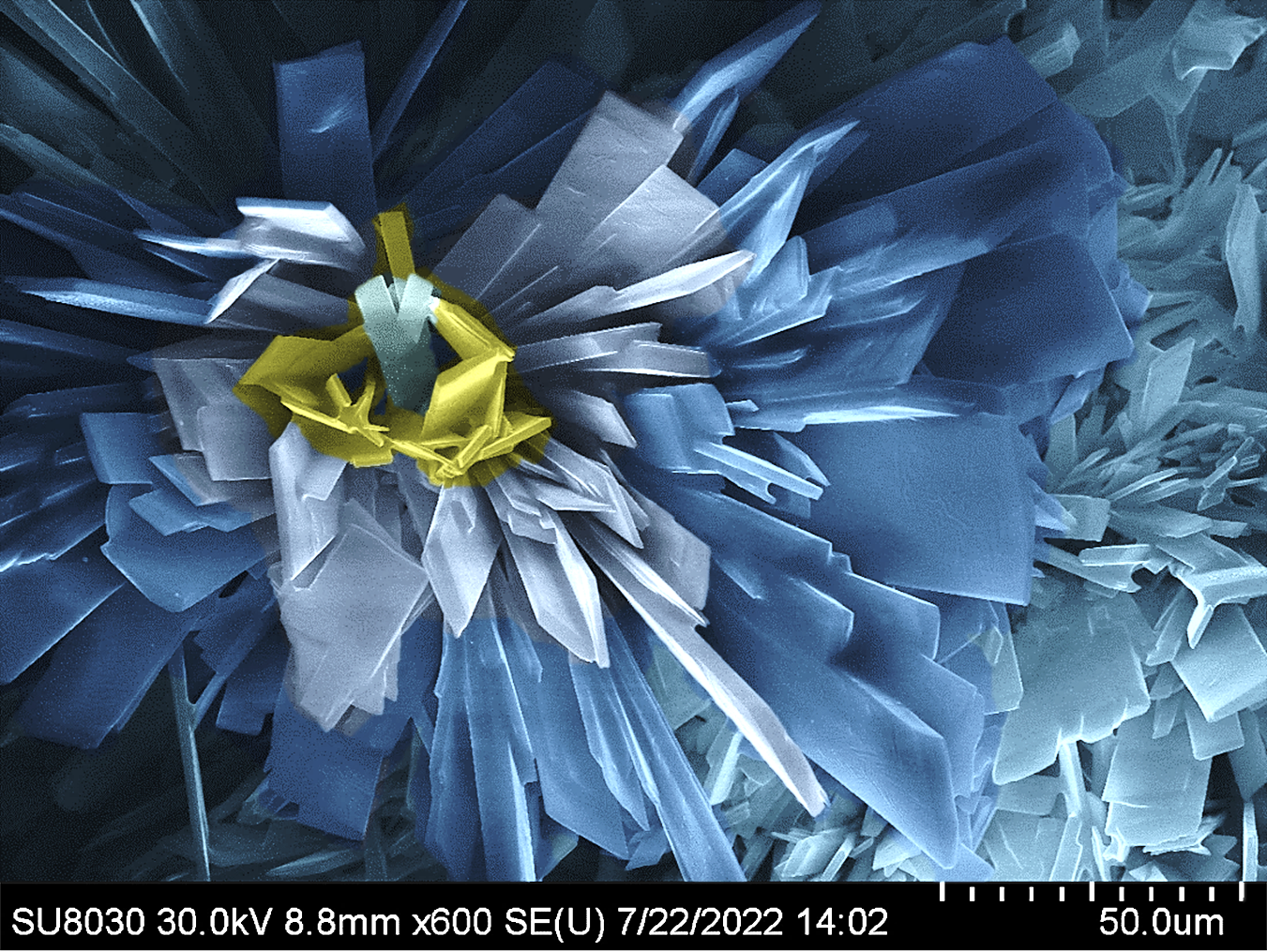
Snowswept Memory
Artist: Haomiao Xie, Post-Doc, Northwestern
NNCI Site: SHyNE
Tool: Hitachi 8030
SEM image of flake-shape Metal-Organic Framework microcrystals.
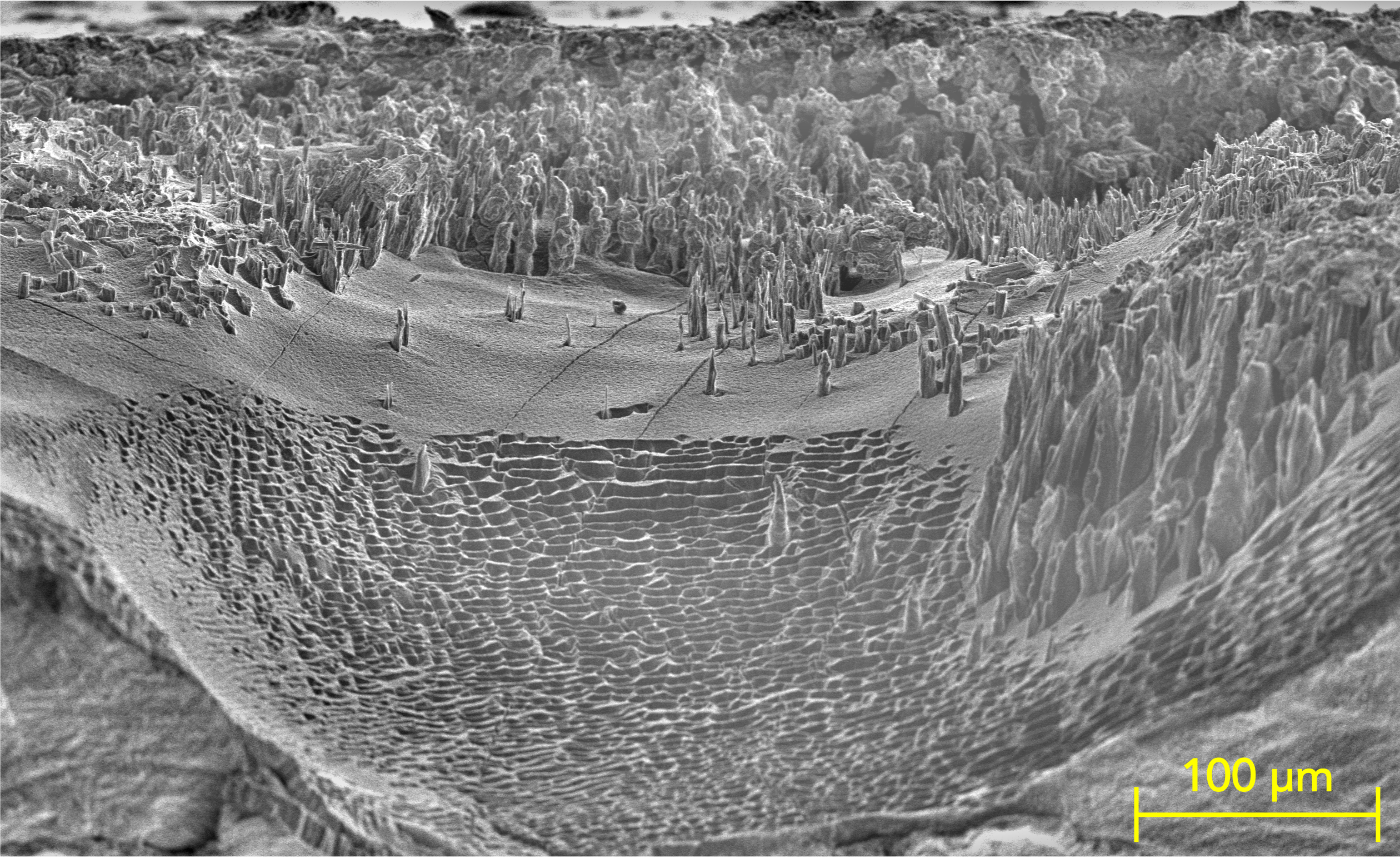
Morning Mist in the Lithium Pine Forest
Artists: Pu Riley Zhang (Graduate Student), Stanford University - Johanna Nelson Weker and Yi Cui Research Groups
NNCI Site: nano@stanford
Tool: SEM: Thermo Fisher Scientific Apreo S LoVac Scanning Electron Microscope
This scanning electron microscopy image was taken after ion milling of the lithium-polymer stack. ImageJ, fit polynomial plug-in, was applied to create the morning mist in this lithium pine forest.
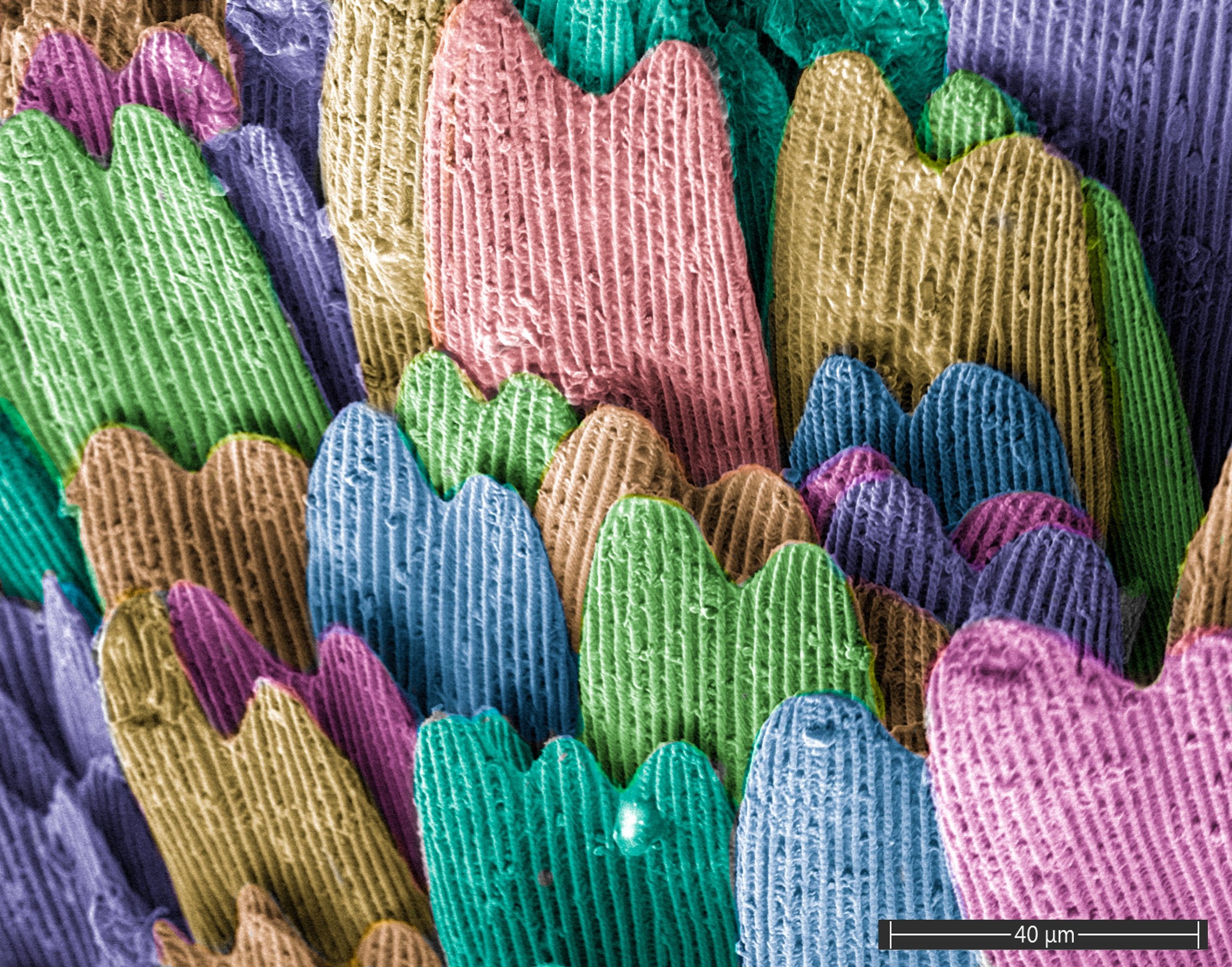
Rainbow Scales
Artist: Evgeniya Moiseeva, MNTC Staff, University of Louisville
NNCI Site: KY Multiscale
Tool: FIE Apreo C SEM
SEM Image of the monarch butterfly wing.
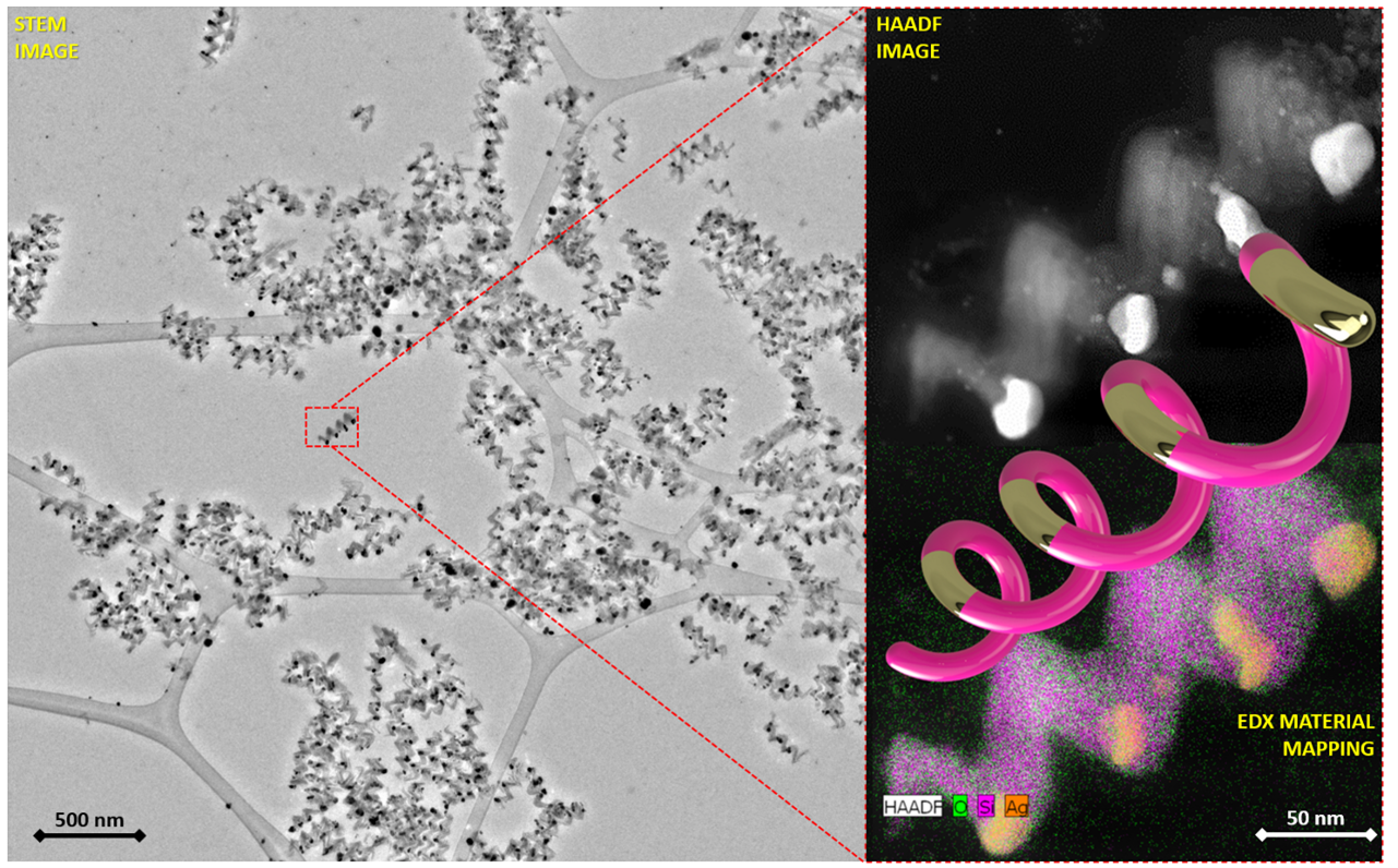
Helical Metamaterial Platform
Artists: Shawn Wimer (Graduate Student), Dr. Ufuk Kilic, and Dr. Matthew Hilfiker, Electrical and Computer Engineering Department, University of Nebraska Lincoln
NNCI Site: Nebraska Nanoscale Facility (NNF)
Tools: FEI Tecnai Osiris (S)TEM
Here, the highly porous, super-lattice type, periodic arrangements of nano-plasmonic, right-handed Silicon (Si)-Silver(Ag) chiral heterostructure metamaterial platform were fabricated by means of subsequent and repeated electron-beam glancing angle deposition from Si and Ag sources. The figure collage shows the scanning transmission electron microscopy (STEM) image which enabled a prominent ability to create contrast and distinguish Si and Ag sub-helical segments (left image), the high-angle-annular-dark-field STEM enabled to focus on isolated single helicaL heterostructure system (right top image) and the atomic resolution energy-dispersive X-ray spectroscopy-based material mapping enables to map the material along single Si-Ag helical heterostruture metamaterial (right bottom image).
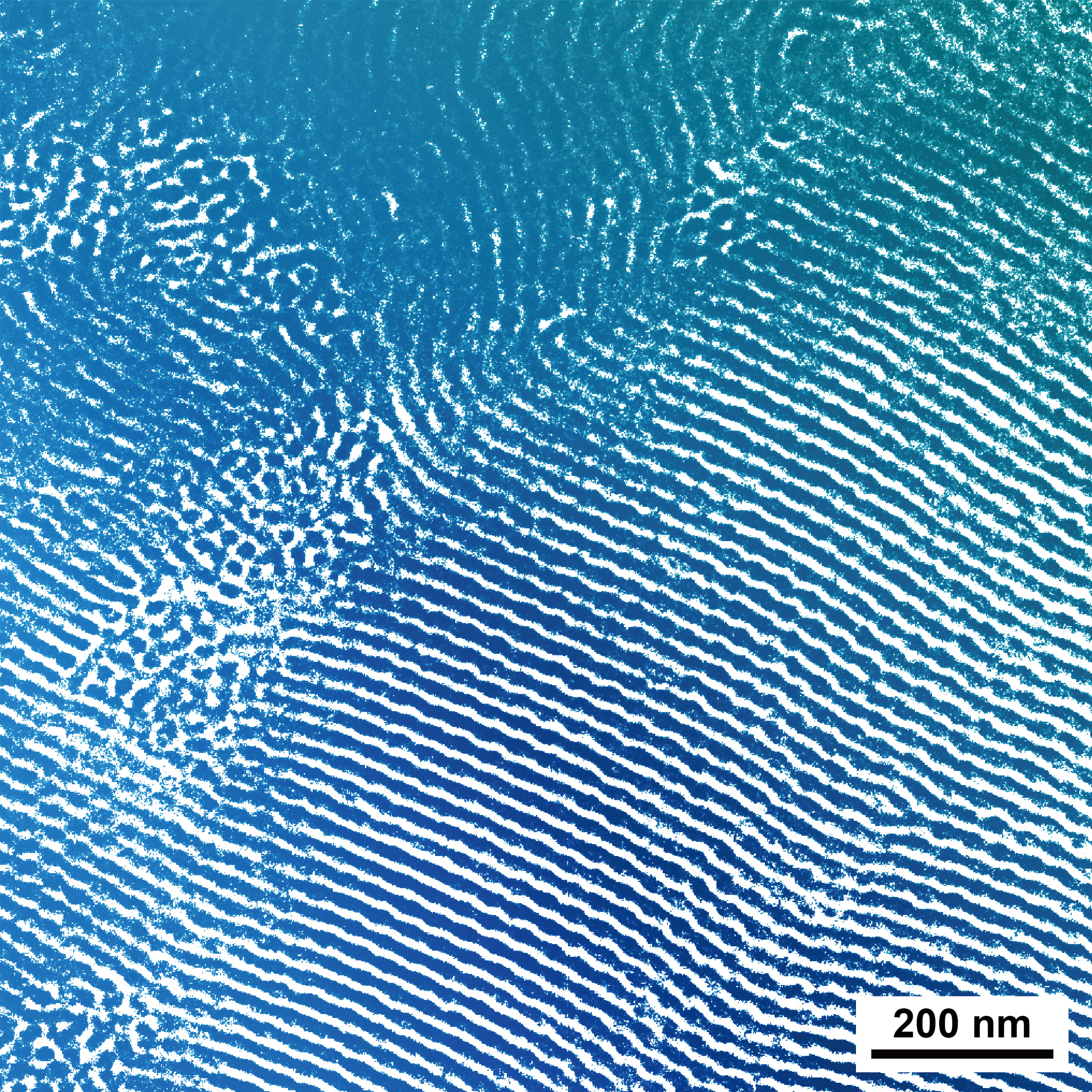
Blue Ocean
Artists: Shuquan Cui (post-doc), Department of Chemistry, University of Minnesota; Liyang Shen (post-doc), Department of Chemical Engineering & Materials Science, University of Minnesota
NNCI Site: MiNIC
Tool: FEI Tecnai Spirit Bio-Twin Transmission Electron Microscope
This stunning blue ocean with vortex and wave is from the self-assembly of block copolymers in the nanoscale. From this image, we can find various amazing structures in the nanoworld, which are just dominated by several simple interactions. Science is like this blue ocean, rich, fascinating, and mysterious, riveting all of us to its beauty.
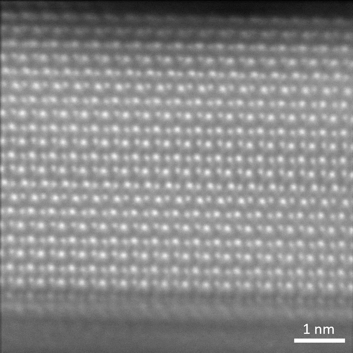
SnSe HAADF-STEM Image
Artists: Jules Gardener, Senior Scientist, Harvard University & Austin Akey, Senior Scientist, Harvard University
NNCI Site: Center for Nanoscale Systems, Harvard University
Tool: TEM-11 JEOL ARM
This high resolution high annular dark field scanning transmission electron microscope (HAADF-STEM) image is of the ferroelectric material SnSe. Alternating bright and dark atoms can be seen, which correspond to Sn and Se respectively, allowing for the direct observation of the crystallographic structure of this novel material. This image was acquired by aligning and summing several raw images, thereby reducing scan noise.
Moon Craters
Artists: David Irvine and Reynolds Dziobek-Garrett, PhD Candidates, Johns Hopkins University
NNCI Site: CNF
Tool: Zeiss Ultra SEM
This image depicts a titled SEM view of vapor-liquid-sold (VLS) growth of silicon under a waveguide ring resonator area before processing it with the chemical mechanical polishing (CMP) machine. The growth method over a large surface area creates a “moon crater” like surface due to the anisotropic catalyzed silicon whisker growth.
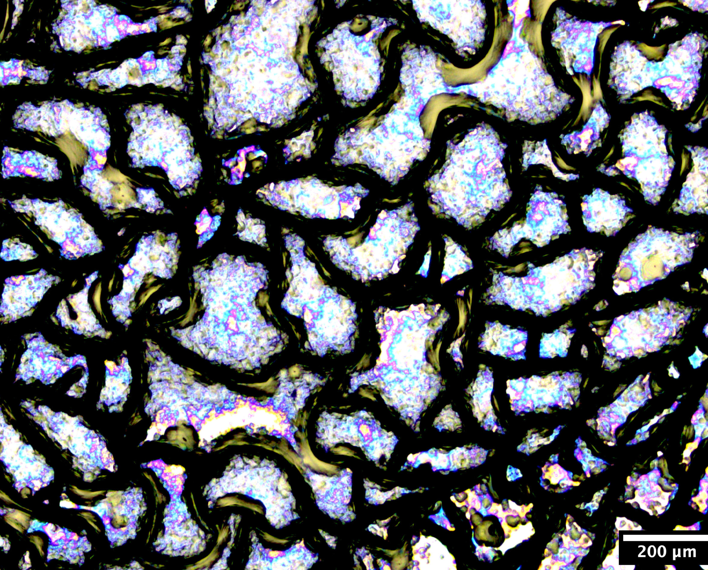
Stained Glass
Artists: Matthew Campbell, Postdoctoral Fellow, University of Pennsylvania and Pawan Kumar, Postdoctoral Fellow, University of Pennsylvania
NNCI Site: MANTH
Tool: Cambridge NanoTech S200 ALD (Al2O3), Denton Vacuum Explorer 14 Sputterer (Mo), SMI MOCVD (MoS2 process)
We are developing highly reflective, lightweight films for laser-powered interstellar travel. One of our prototypes became corroded during one of the fabrication steps and looked like this in the optical microscope. More specifically, we grew a thin metalized film on top of a ceramic film that coated a double-side-polished silicon chip. The backside of the chip was corroded during the growth process of the metalized film. We deposited another thin ceramic film over the thin metalized film.
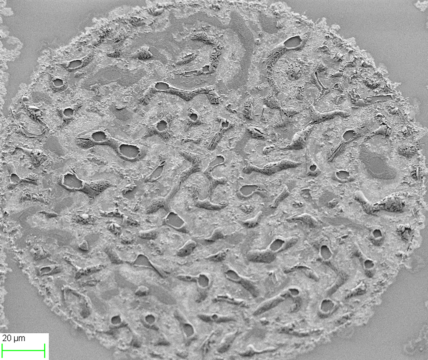
Beautiful Mistake
Artists: Aaron Bell (Bio Electron Microscopy Staff Scientist) & Jin Nakashima (Senior Research Scholar), NC State University
NNCI Site: Research Triangle Nanotechnology Network (RTNN)
Tool: Hitachi HT7800 TEM
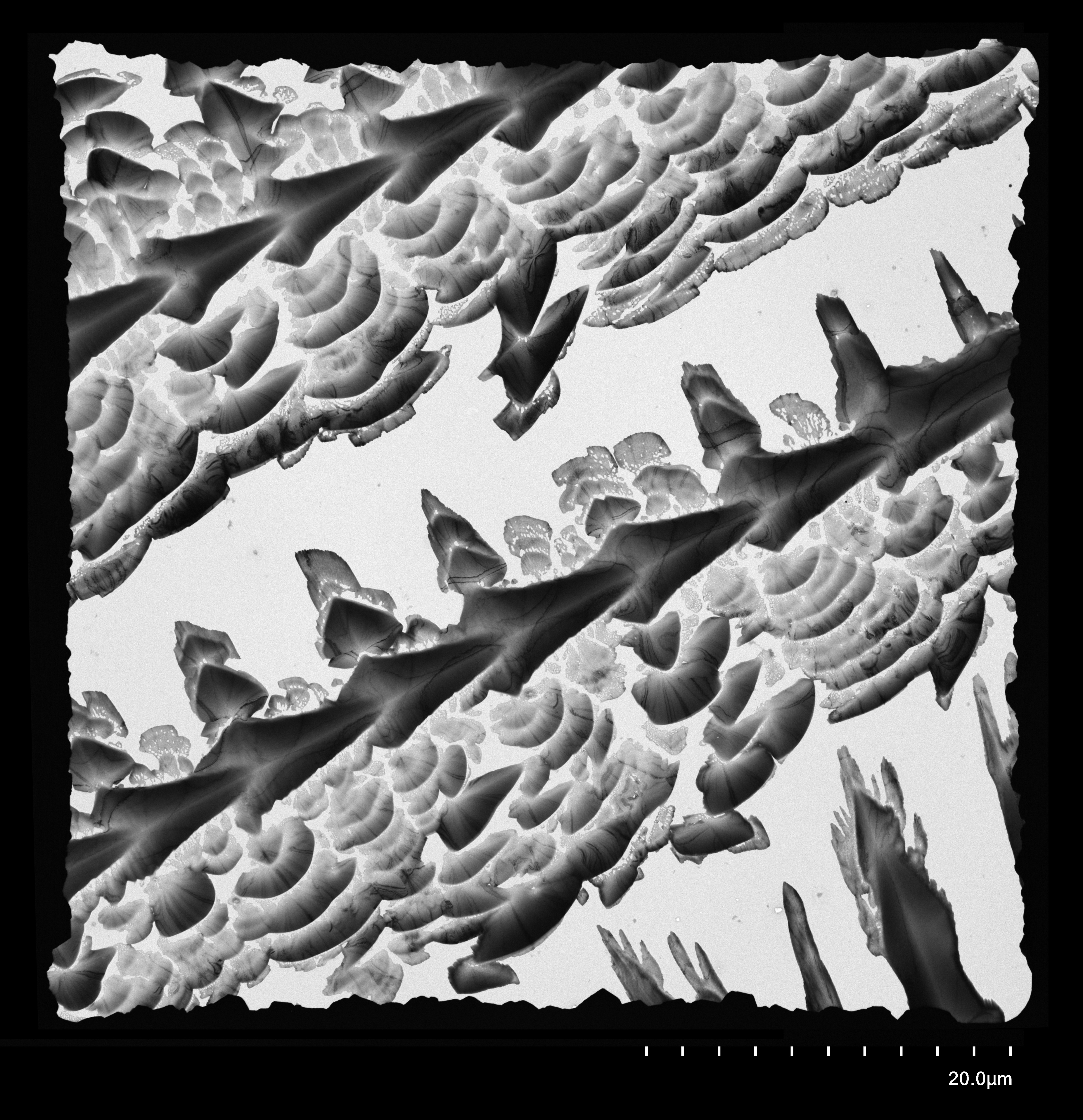
This was a negative stain that went wrong (but “oh so right”). We're guessing that there was something in the buffer that made the uranyl acetate precipitate into these amazing shapes. The specimen itself was unusable for scientific purposes but the images themselves were quite striking.

Micro-Trusses
Artist: Zainab Patel, Graduate Student, University of Washington, Materials Science & Engineering
NNCI Site: Northwest Nanotechnology Infrastructure (NNI)
Tool: SEM Apreo in the UW Molecular Analysis Facility
Damage resist truss made of two interpenetrating lattices made using two-photon lithography aka nanoscale 3D printing.
Lava Dew Drops on Obsidian Grass Blades
Artists: Mohammad Bakhtiar Uddin (PhD candidate) and Dr. Ram V. Mohan, North Carolina A&T State University/Joint School of Nanoscience and Nanoengineering
NNCI Site: SENIC
Tool: Zeiss Auriga FIB/FESEM
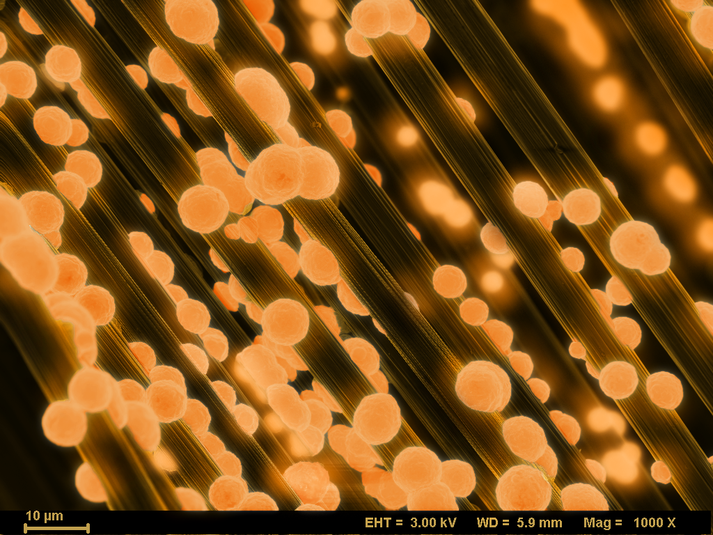
Metallic copper droplets were deposited on carbon fiber tow through electroplating to create this pattern. Polymer coating on the surface of the carbon fiber prevents the fiber from being wetted by copper which creates bead like droplets depicted in the image.
Christmas Trees
Artists: Penghui Zhao, Postdoc and Wujin Sun, Assistant Professor, Biological Systems Engineering
NNCI Site: NanoEarth
Tool: JEOL IT500HR Scanning Electron Microscope
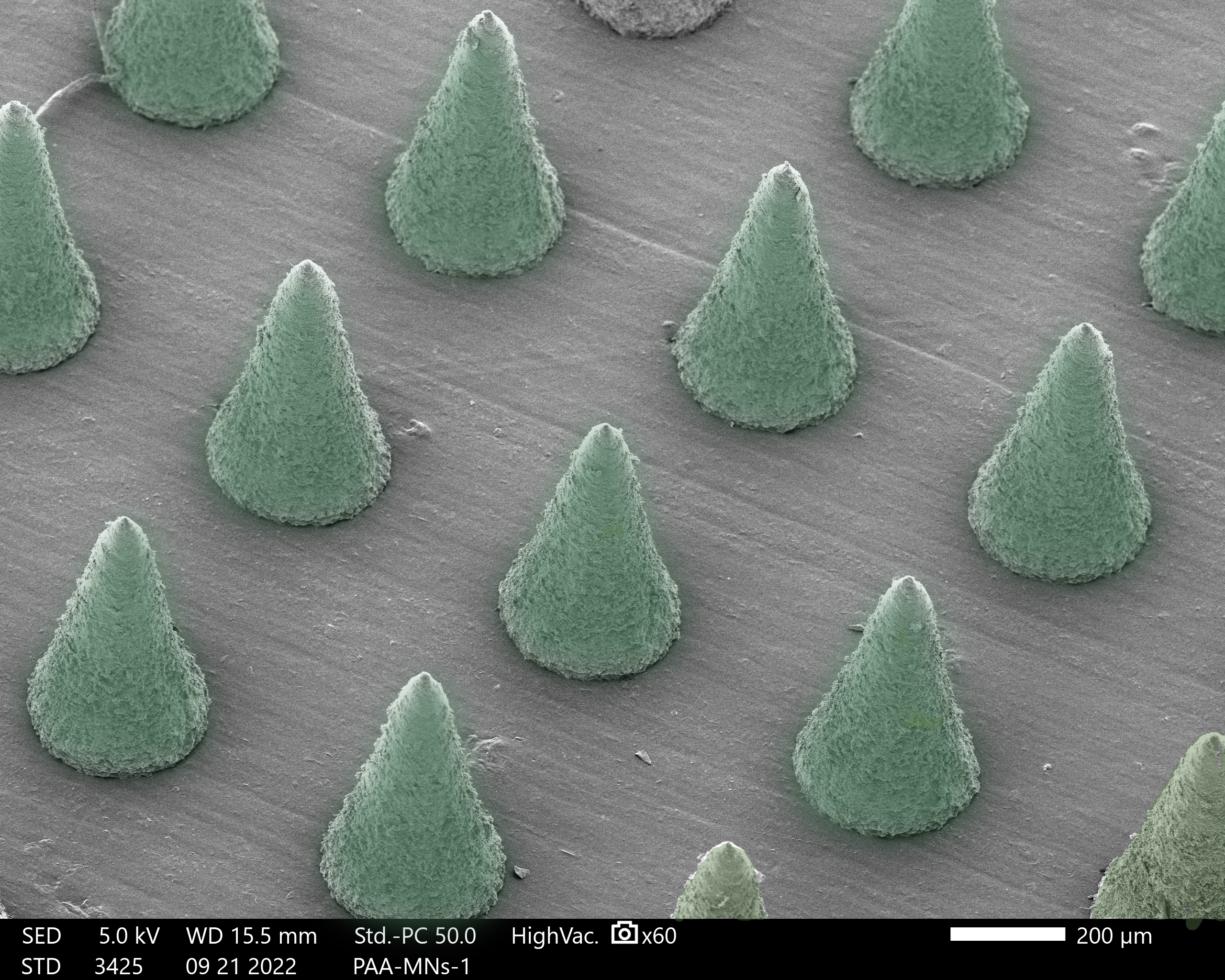
Christmas trees bring happiness to thousands of families, as our microneedles bring health to a large majority of people in the world. Microneedles provide an appealing administration approach to patients as it's painless, convenient, and self-administered. This featured SEM image of microneedle is fabricated by polyacrylic acid, it has a brilliant dissolvable ability, The microneedles could penetrate into the targeted area, release drugs, and drive away disease. As happiness and warmth emerging in Christmas Eve of thousands of households, carried by Christmas trees.