
NNCI Image Contest 2021 - Whimsical
Most Whimsical
Artists in this category playfully used micro and nanoscale images as the foundation to build scenes. Please check out the images below and read a little about the research behind them.
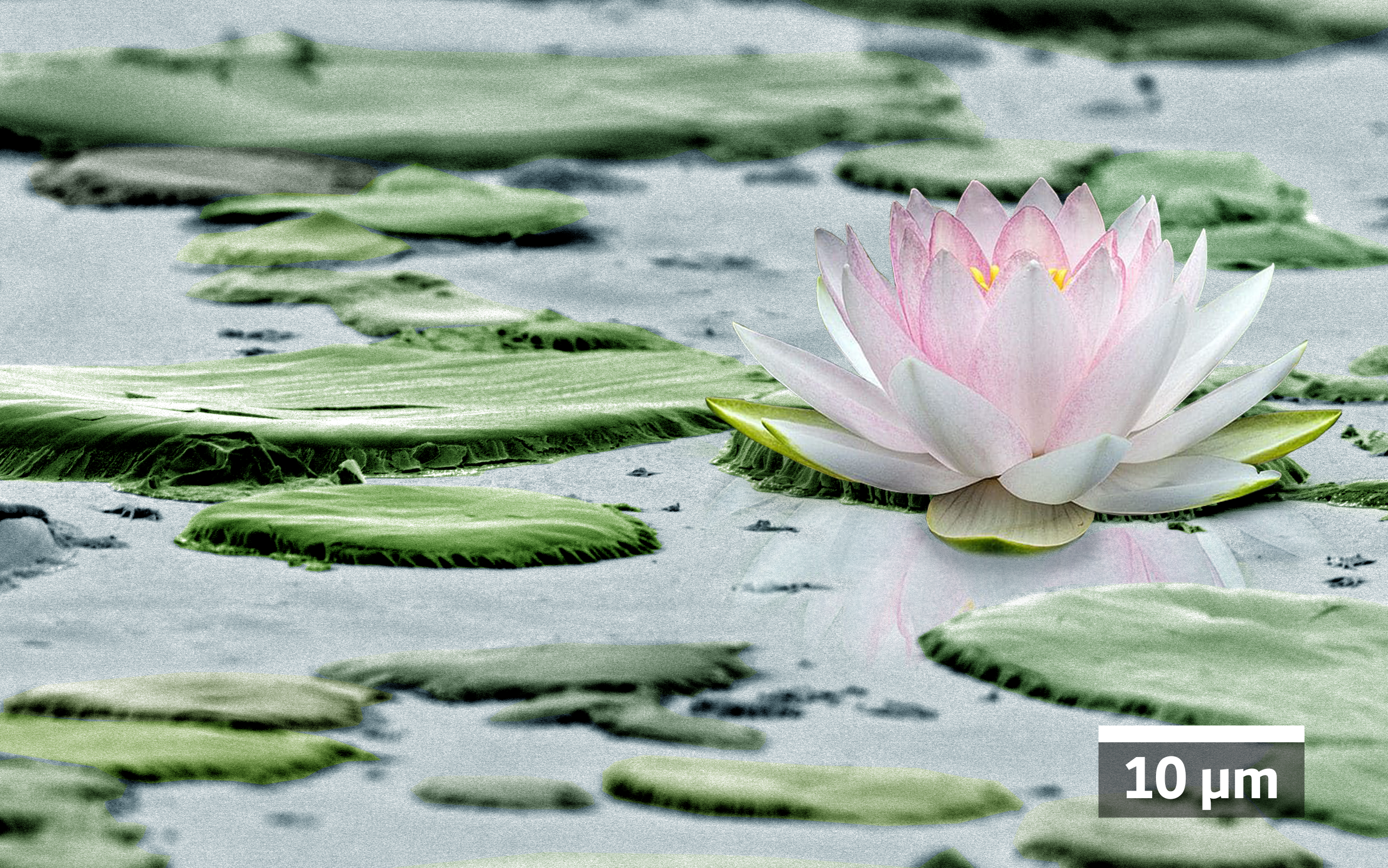
Lotus on Anti-SARS-CoV-2 Coating
Artists: Mohsen Hosseini, Ph.D. Student and William A. Ducker, Professor, Virginia Tech
NNCI Site: NanoEarth
Tool: JEOL IT500HR Scanning Electron Microscope
Transparent silver based antimicrobial coating. The coating is a thin film made out of particles bound to glass substrate and has been specifically made as our response to the COVID-19 pandemic as engineers. One of the transmission routes for SARS-CoV-2 is through contact with contaminated objects and touching nose or mouth accordingly. This coating limits this transmission route. The coating has also demonstrated the ability to kill bacteria such as P. aeruginosa, S. aureus and MRSA. The scanning electron microscopic image shows the beauty at the micro scale of this coating, implying a lotus flower (nelumbo nucifera) environment.
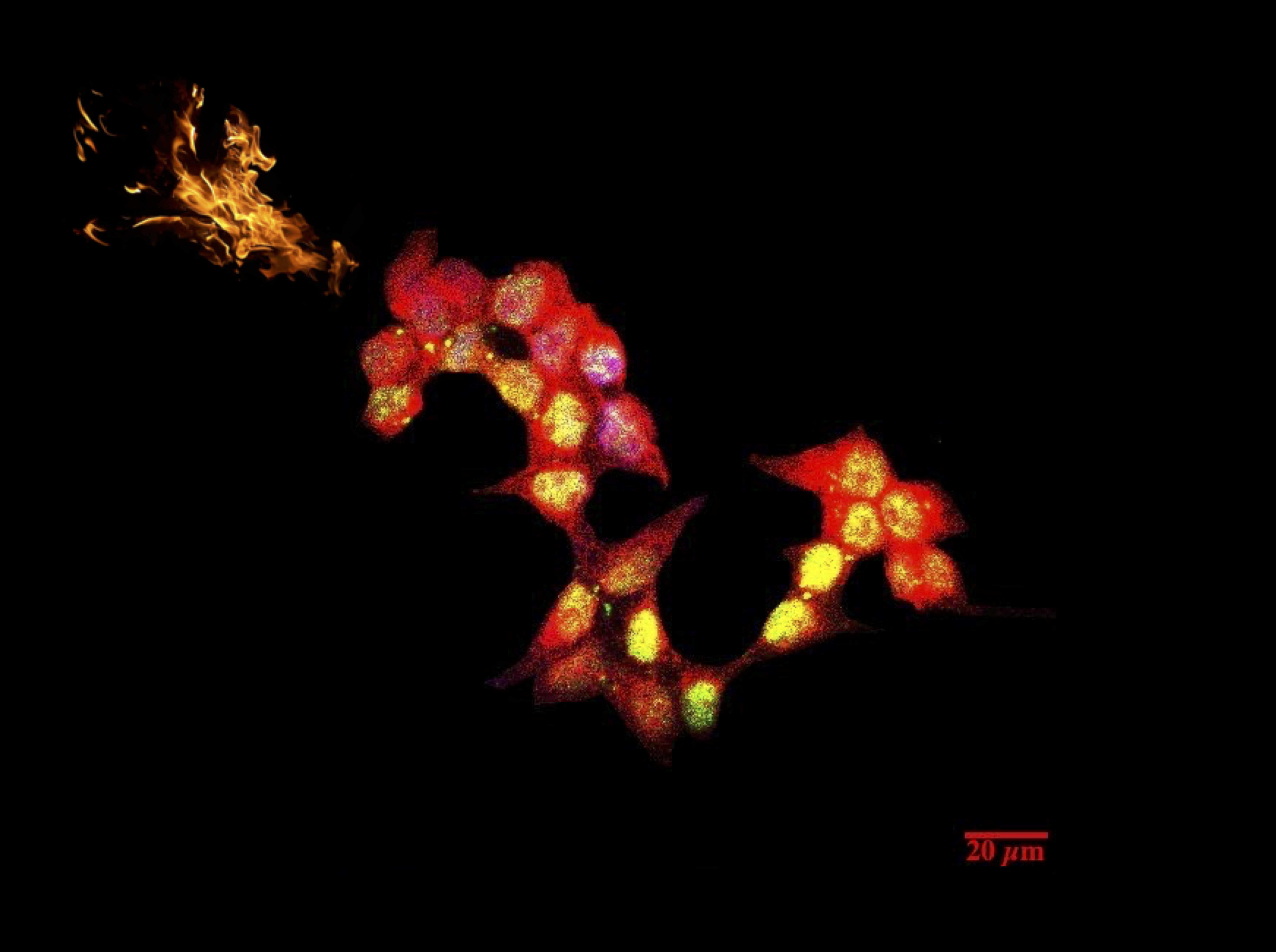
Dragon HEK293T cells
Artist: Mona Motaghed, PhD candidate (Graduate student), The Joint School of Nanoscience and Nanoengineering (JSNN)
NNCI Site: SENIC
Tool: Zeiss AXIO Observer Z1 Spinning Disc Confocal Microscopy
Imagine that you have an unlimited source of your favorite immune cells as a trojan horse to combat many cancers. To achieve this goal, we are using HEK293T cells to produce the lentiviruses carrying the GOI. Then, the lentiviruses will be used to reprogram human Mast Cells to combat different cancers. HEK293T cells stably express the SV40T-Antigen which can bind to SV40 enhancers of expression lentivirus vectors to increase protein production. Briefly, HEK293T cells were incubated with Mouse-SV40T Antigen primary antibody and then stained with Donkey-Anti Mouse IgG-FITC secondary antibody. The mitochondria and nucleolus were stained using Mitotracker-Red and DAPI, respectively.
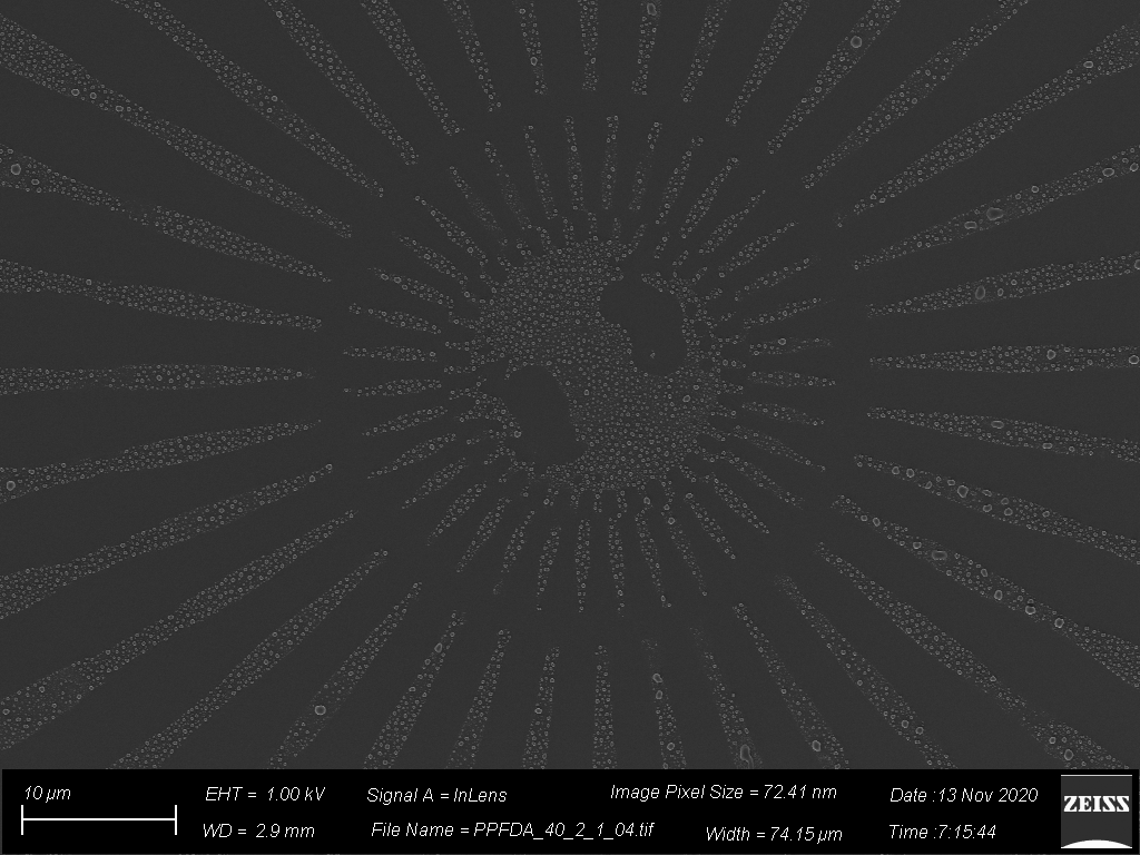
World of Islands
Artists: Dani Streever, undergraduate, and Trevor Donadt, PhD candidate, at Cornell University
NNCI Site: CNF
Tool: SEM & iCVD reactor & lithography
Droplets of polymer were deposited onto a microscale etched pattern. The resist of the pattern was then dissolved away and the droplets were left over in the shape of the pattern. This was part of a project to show proof of concept for patterning and controlling polymer depositions on a nanoscale..
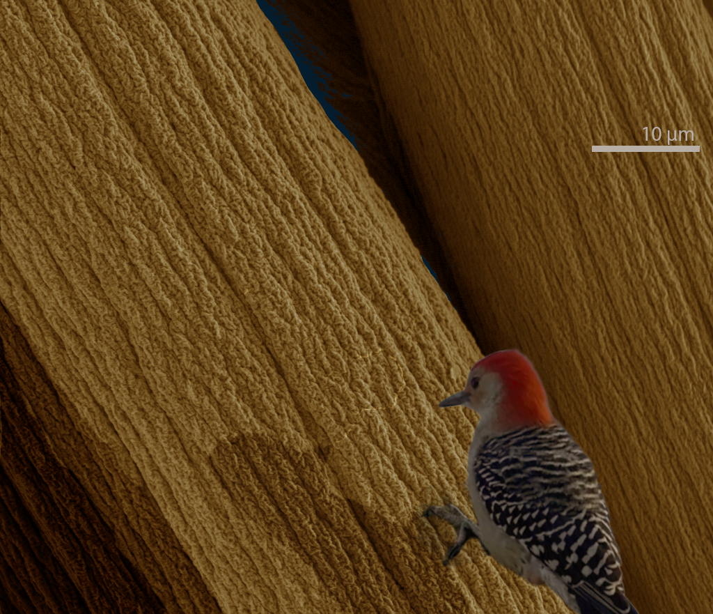
Arboreal Alginate
Artists: Christopher Johnson, PhD Candidate, and Ravisara Wattana, PhD Student, University of Pennsylvania
NNCI Site: MANTH
Tool: FEI Quanta 600 ESEM
This is a scanning electron microscope image of an alginate fiber, one of Earth's most abundant biomaterials, obtained from brown algae. Alginate is biocompatible, biodegradable, and a renewable resource for applications such as biomedical scaffolds or rheological studies. Our research is focused on optimizing conditions for the formation of these fibers in both mechanical and chemical contexts, and then observing their material properties. The fibers in this image were formed by extruding into a calcium chloride bath. Without the false color presented in this image, alginate fibers are typically clear and flexible, ranging from dozens to hundreds of microns thick.
A Summer Snack for NanoAnts
Artist: Eric Roth, BioCryo Electron Microscopy Specialist, NUANCE Center, Northwestern University
NNCI Site: SHyNE
Tool: Hitachi HD2300 STEM
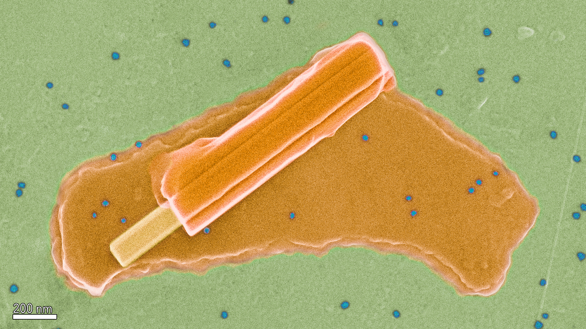
This fun feature was found on a grid used for training researchers to use the Hitachi HD2300 STEM. The popsicle and stick are made from a molybdenum crystal, the melted part is NaCl, and the, "ants" are 25 nm gold nano particles.
Rocky Marshland
Artist: Oghenetega Allen Obewhere, Grad. Student, University of Nebraska-Lincoln
NNCI Site: NNF
Tool: Anasys afm+ AFM
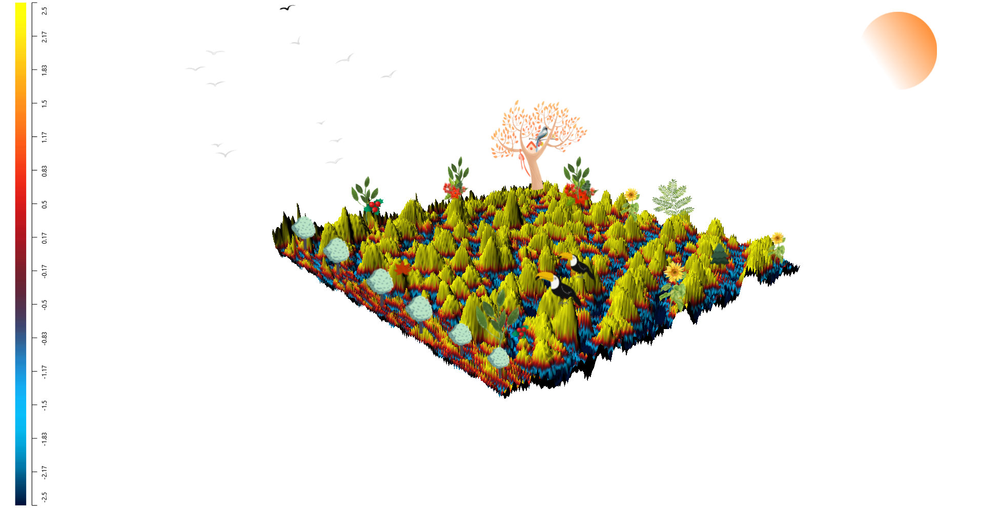
A 3-D contact resonance atomic force microscopy (CR-AFM) phase image of thin-film ion-conducting polymer (Nafion) and Platinum nanoparticles (Pt-NPs). Ion conducting polymers play a crucial role as binders on the electrode surface of electrochemical devices where they help to fix the catalysts to the electrode surface. An optimized ratio of catalyst/ionomer is needed for the efficient electrochemical reactions at the electrodes of these devices. This CR-AFM surface image elucidates the extent of nanoparticles dispersion within the ionomer film (Nafion, blue and Pt-NPs, green) at this loading and it has been recreated as a rocky coastal marshland in the dark.
Dinos Take the City
Artist: James Loveless, Graduate student, North Carolina State University
NNCI Site: RTNN
Tool: Field Emission Scanning Electron Microscope - FEI Verios 460L
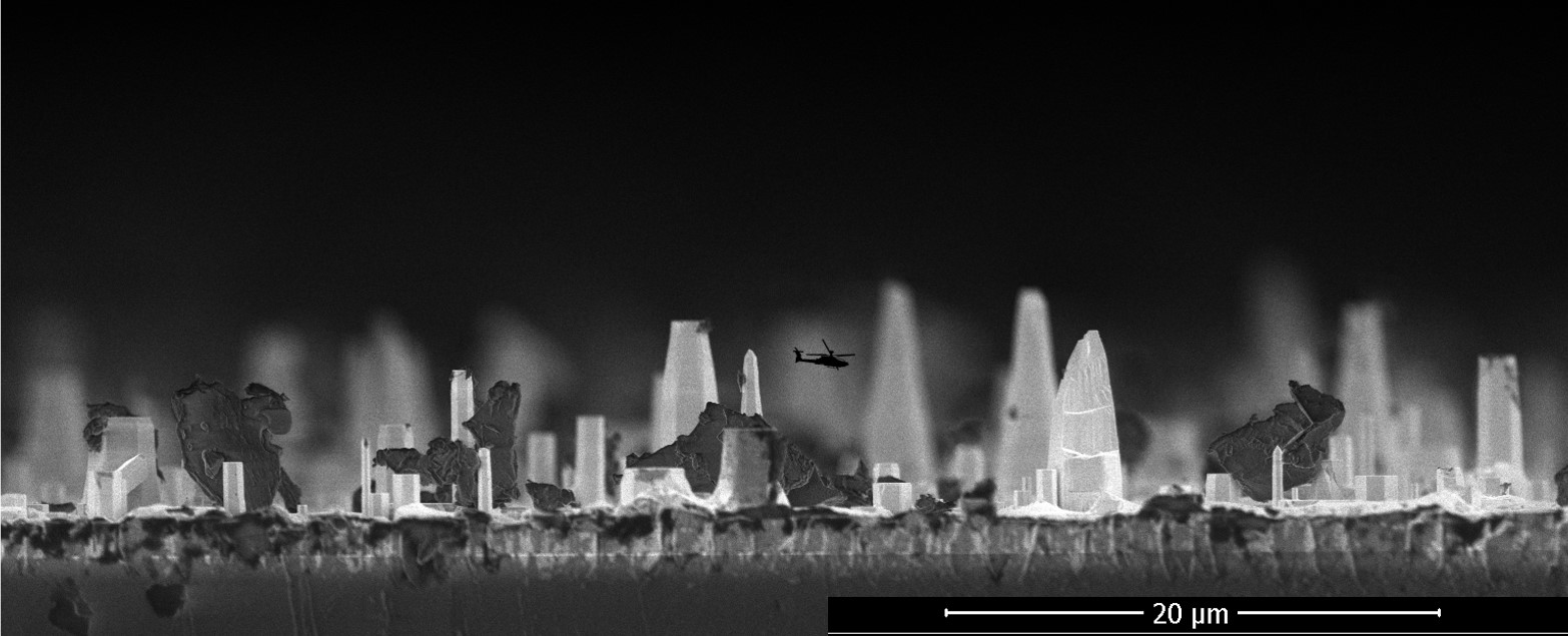
This sample is Gallium nitride grown on sapphire at very high temperatures. Remarkably, this surface was intended to be flat! At these extreme conditions, the crystal growth is often three-dimensional, and we discovered a city-like landscape of thin crystalline microstructures. I thought the dust particles looked like monsters akin to Godzilla, so I added a helicopter to help protect the city.
Micro Bouldering
Artist: Maja Bachmann, Postdoctoral Fellow, Applied Physics, Ian Fisher Research Group, Stanford University
NNCI Site: nano@stanford
Tool: SEM/FIB: FEI Helios NanoLab 600i DualBeam
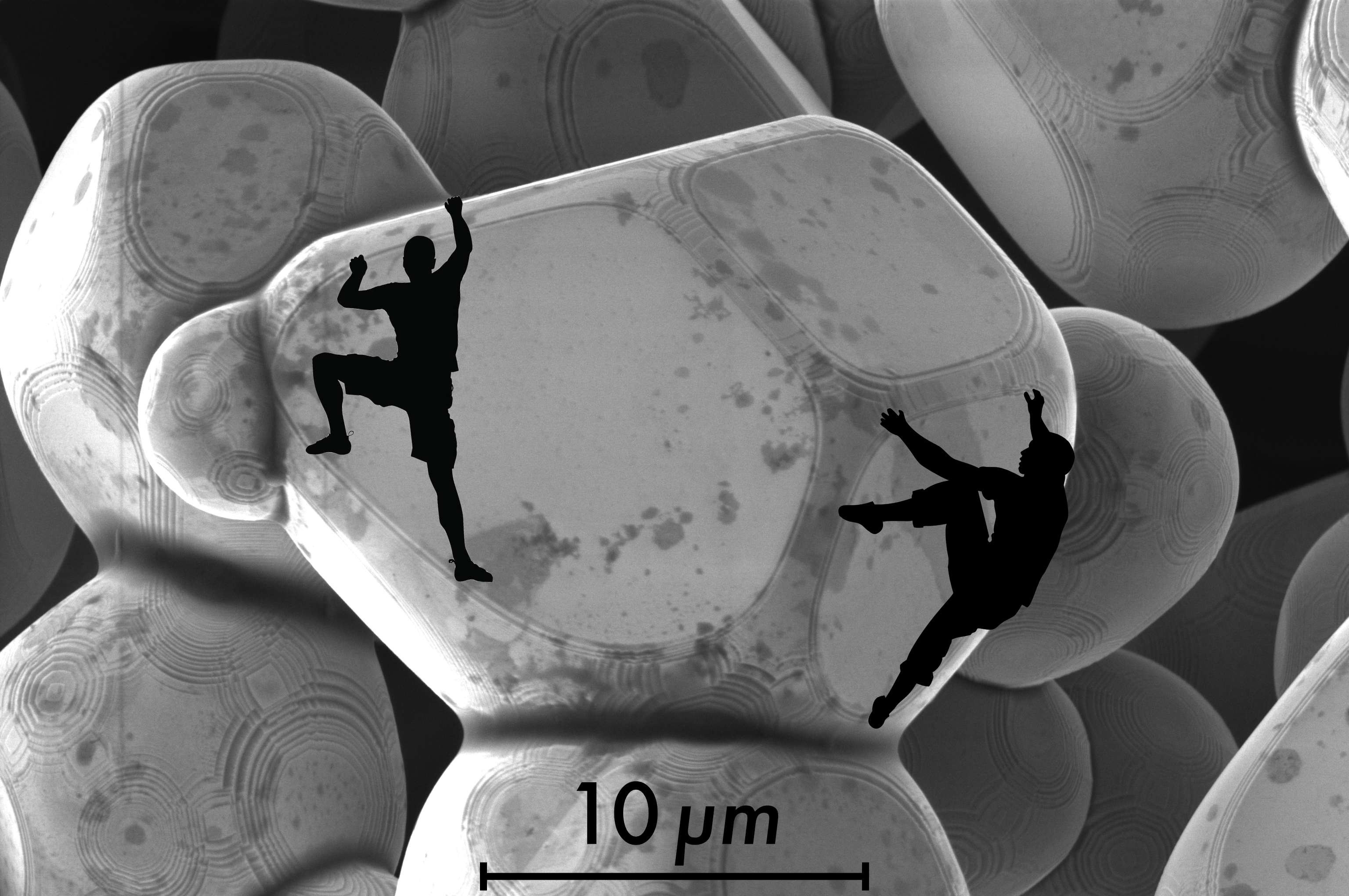
Who wouldn’t want to go bouldering in between these beautiful NiO crystals? The natural step edges follow the symmetry of the underlying crystal structure and would make for perfect crimps and pinches. The crystals displayed here were synthesized at 1400°C using a flux growth technique. Too bad they are only tens of microns large, such that their intricate details can only be marveled at through a SEM and the prospect of bouldering is left to our imagination.
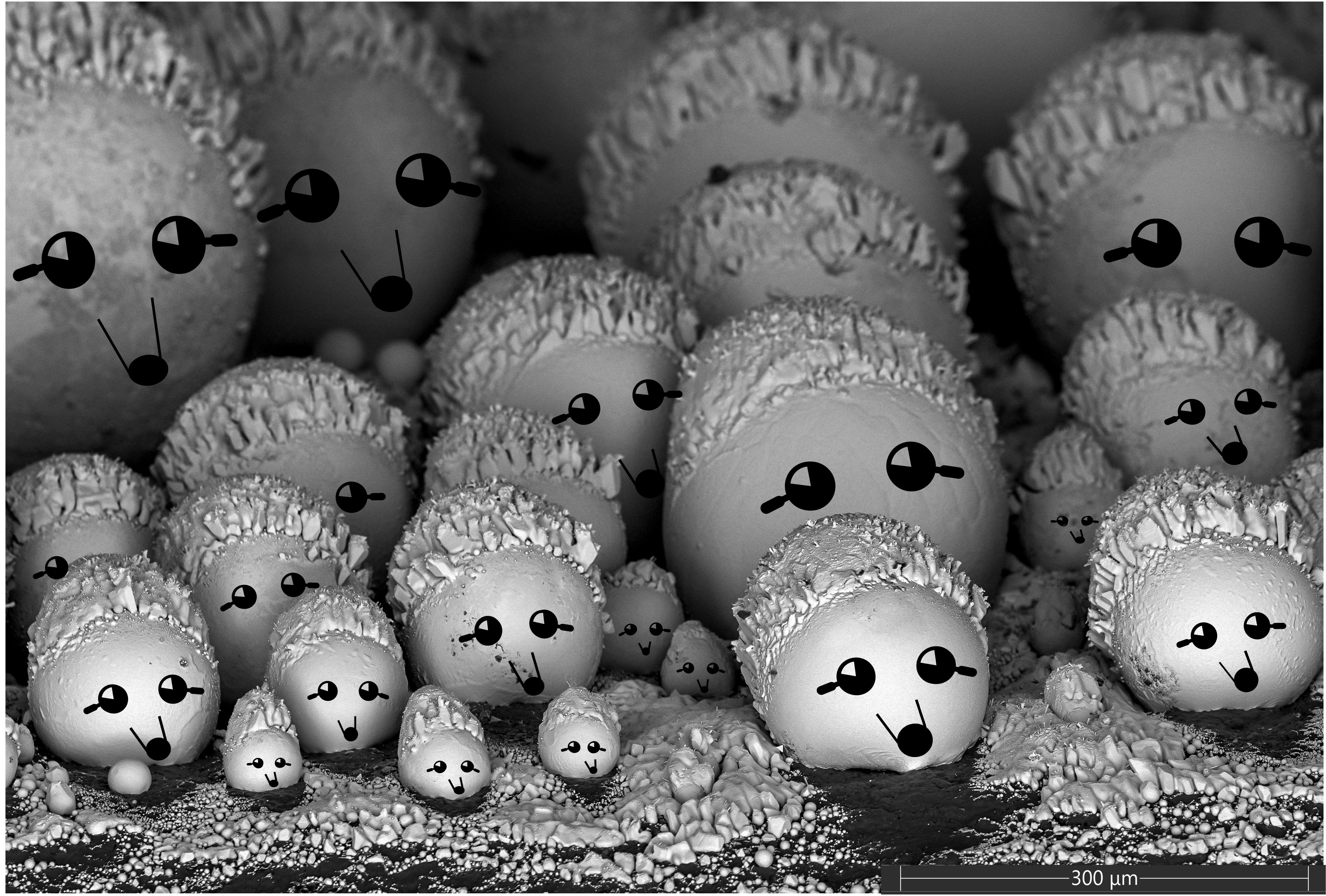
Hedgehog Crew
Artist: Evgeniya Moiseeva, Huson Lab, University of Louisville
NNCI Site: KY Multiscale
Tool: Thermo Scientific Apreo C SEM
Solidified droplets of Gold on the side of the E-Beam evaporation ceramic crucible.
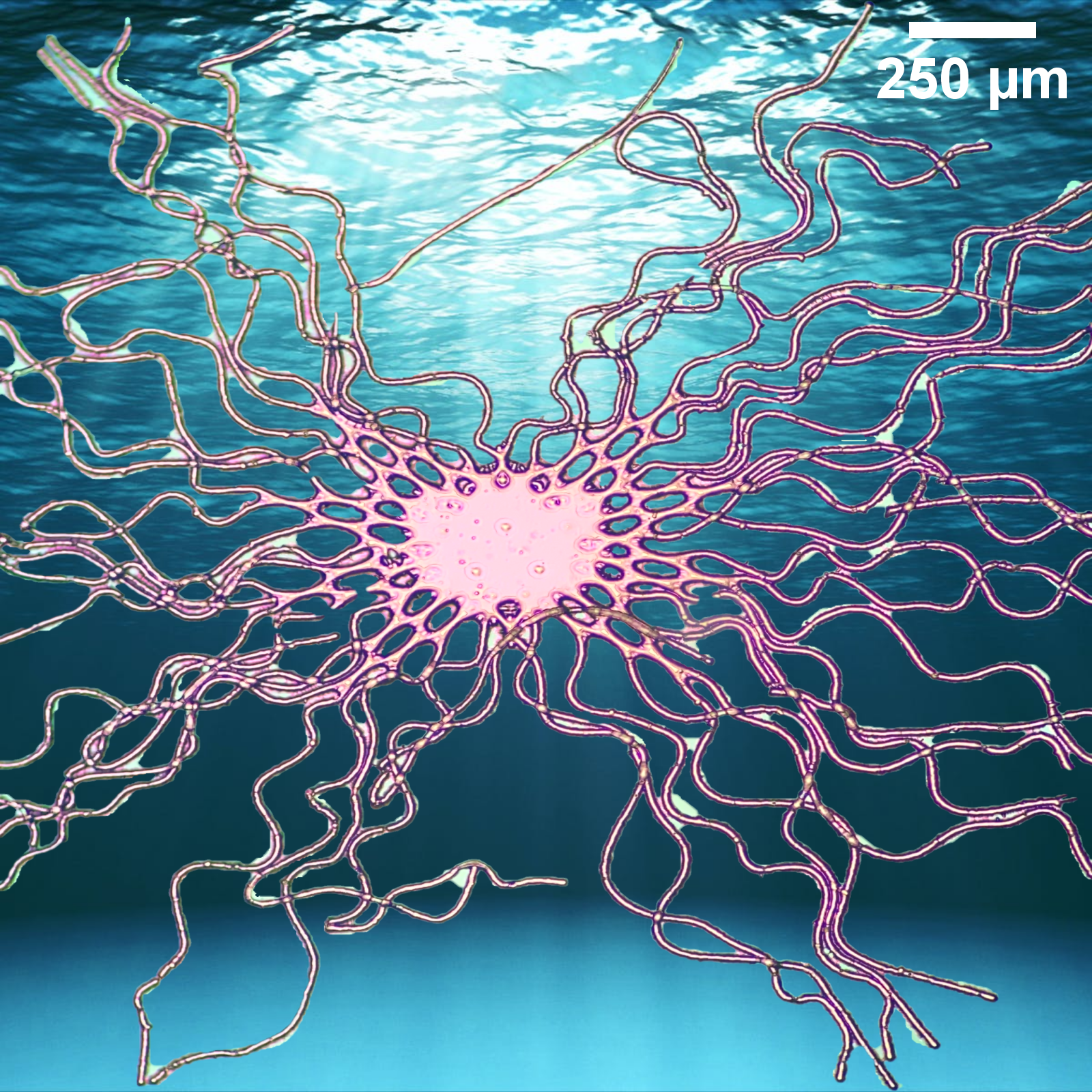
Nanoctopus
Artist: Mackenna Landis, Graduate student, Montana State University
NNCI Site: MONT
Tool: Nikon LV150 Brightfield & Darkfield Reflected Light Microscope
Failed Photoresist! KMPR photoresist was exposed under a mask which contained an array of straight lines to form microchannels using soft lithography. Unfortunately, the resist was accidentally spun on a silicon dioxide-treated wafer, so it did not adhere well. While developing the features, the wafer was placed on a rotating plate, and the unadhered lines of resist began to wobble. Fortunately, this pattern turned out beautifully, if not functional.
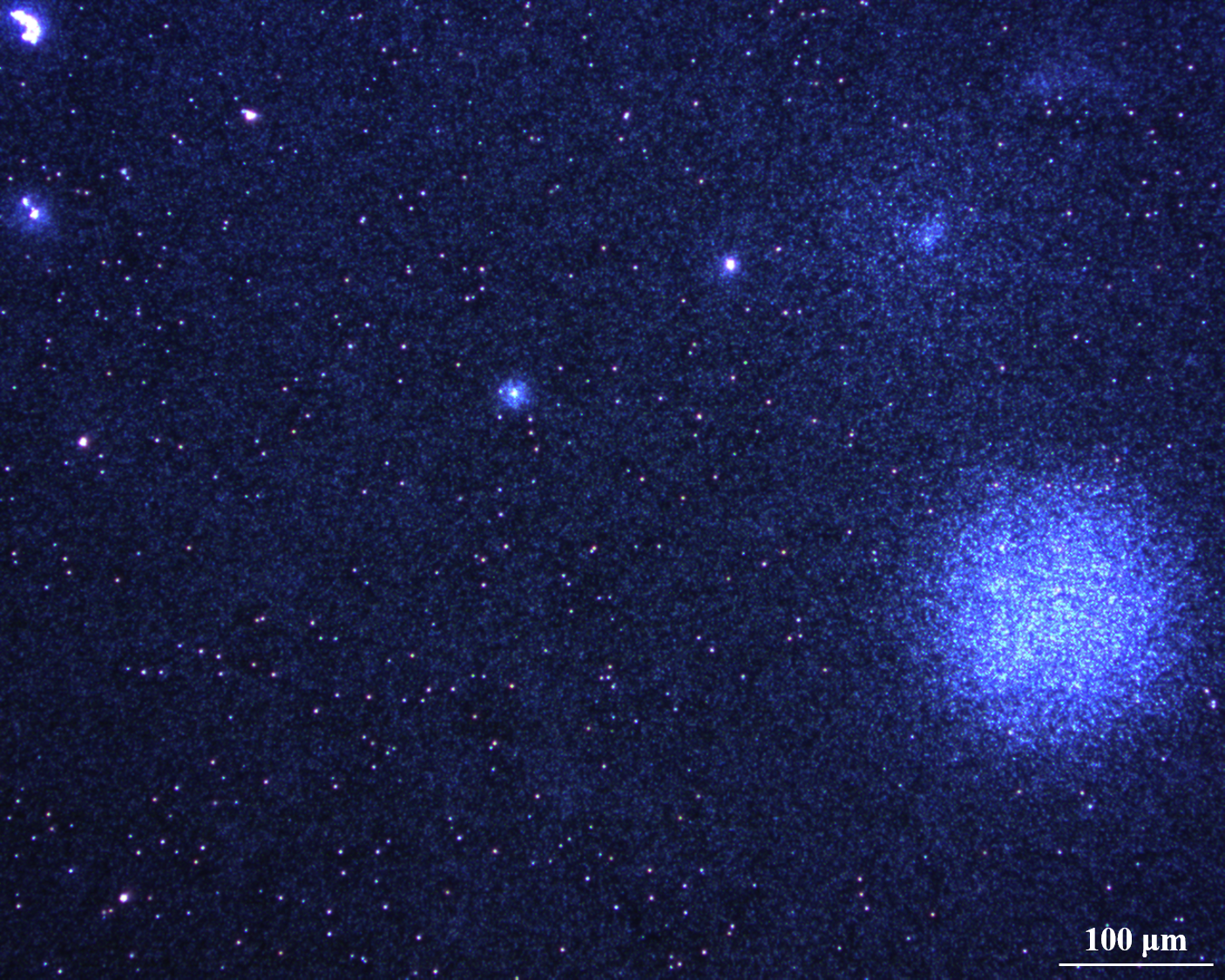
Micro-Nano Starry Sky
Artists: Zihao Lin, Graduate student, and Jeong-Hyun Cho, Faculty member, Department of Electrical and Computer Engineering, University of Minnesota, Twin Cities
NNCI Site: MiNIC
Tool: No tools intentioned to use, captured by Nikon ECLIPSE LV 150
Art in science is often discovered unintentionally. No tools and no design intentioned for this image. We aimed to fabricate devices via a series of fabrication procedures, but darkfield mode in the microscope was used by mistake to observe the sample after several procedures. Surprisingly, we observed this on the silicon chip and took the image. This is a micro-nano starry sky.
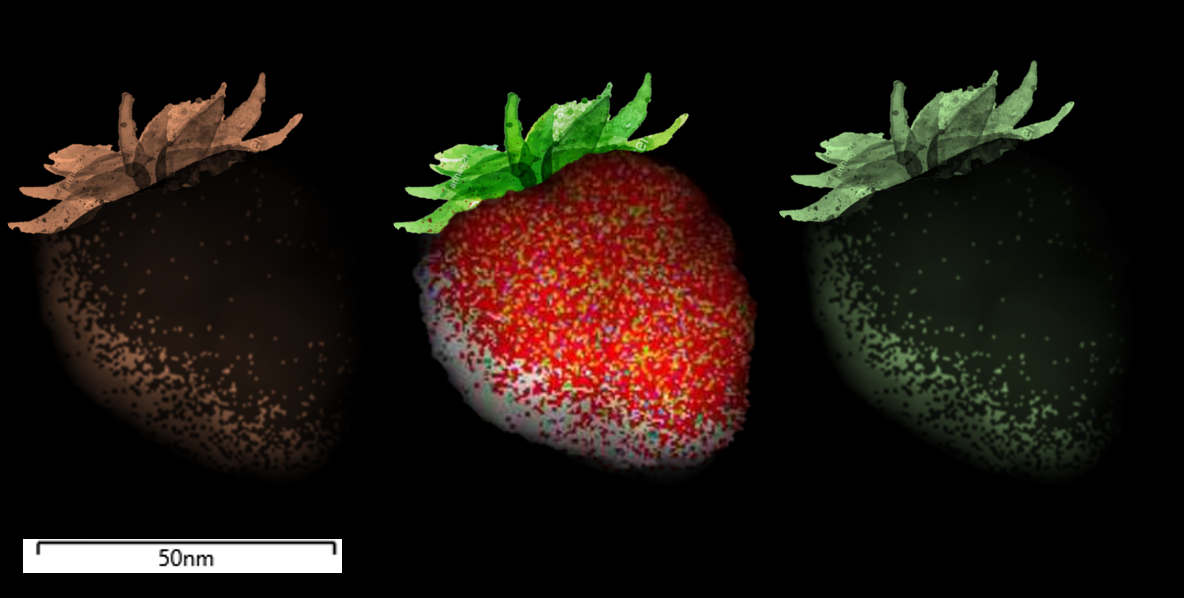
Nano Strawberry Fields Forever
Artists: Ana C. Barrios, Graduate Student, and Francois Perreault, Assistant Professor, Arizona State University
NNCI Site: NCI-SW
Tool: JEOL TEM/STEM ARM200F
Who knew nanoparticles could be this tasty? Don’t let these strawberries fool you! This is an image of a silver nanoparticle (center) that was used to remove bromide from synthetic surface waters. The energy dispersive X-ray microanalysis shows multiple elements including bromide (blue), chloride (green), sulfur (magenta), and phosphorous (yellow) interacting with silver (red). The original image with elemental mapping was not altered. Duplicates of the original image were colored, and leaves were added to enhance the strawberry illusion.
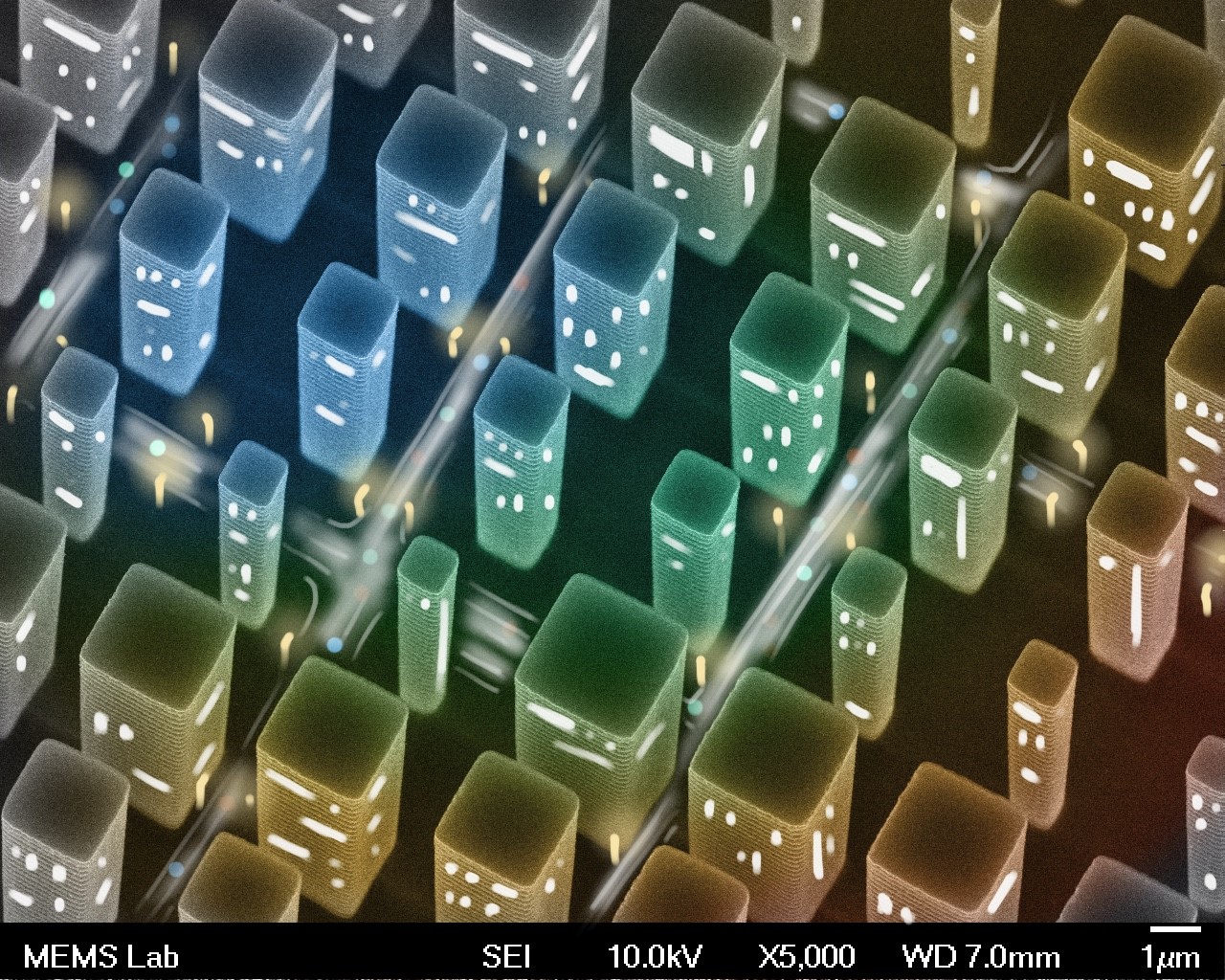
Micro-City
Artists: Zheyi Han, Graduate Student, and Luocheng Huang, University of Washington
NNCI Site: NNI
Tool: JEOL-JSM7400F Scanning Electron Microscope
False-colored and decorated scanning electron microscopic image capturing an array of square silicon pillars from an oblique perspective.