
NNCI Image Contest 2021 - Stunning
Most Stunning
This category celebrates the beauty of the micro and nanoscale. Please check out the images below and read a little about the research behind them.
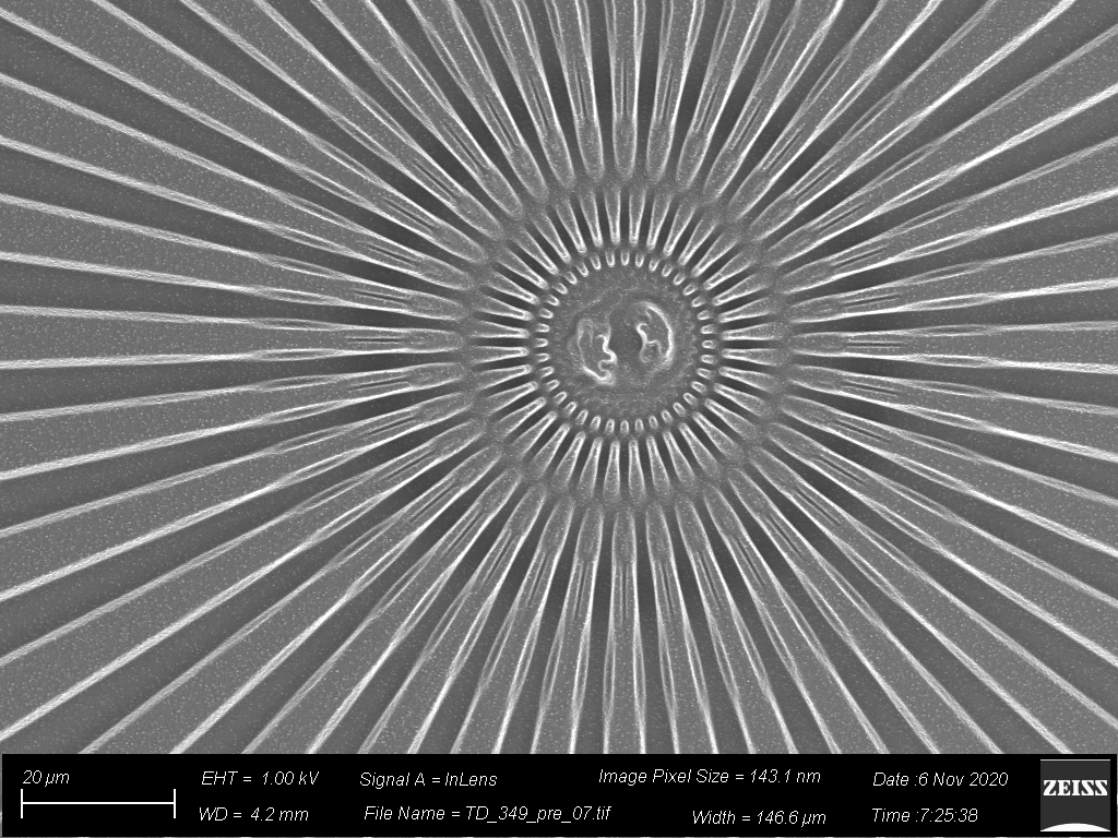
Ring of Spears
Artists: Dani Streever, undergraduate, and Trevor Donadt, PhD candidate, at Cornell University
NNCI Site: CNF
Tool: SEM & iCVD reactor & lithography
A layer of the polymer, PFDA, was deposited onto a microscale etched pattern. This was the first step in part of a project to show proof of concept for patterning and controlling polymer depositions on a nanoscale. The thin film deposition of the polymer was able to well coat the pattern while retaining the microscale details.
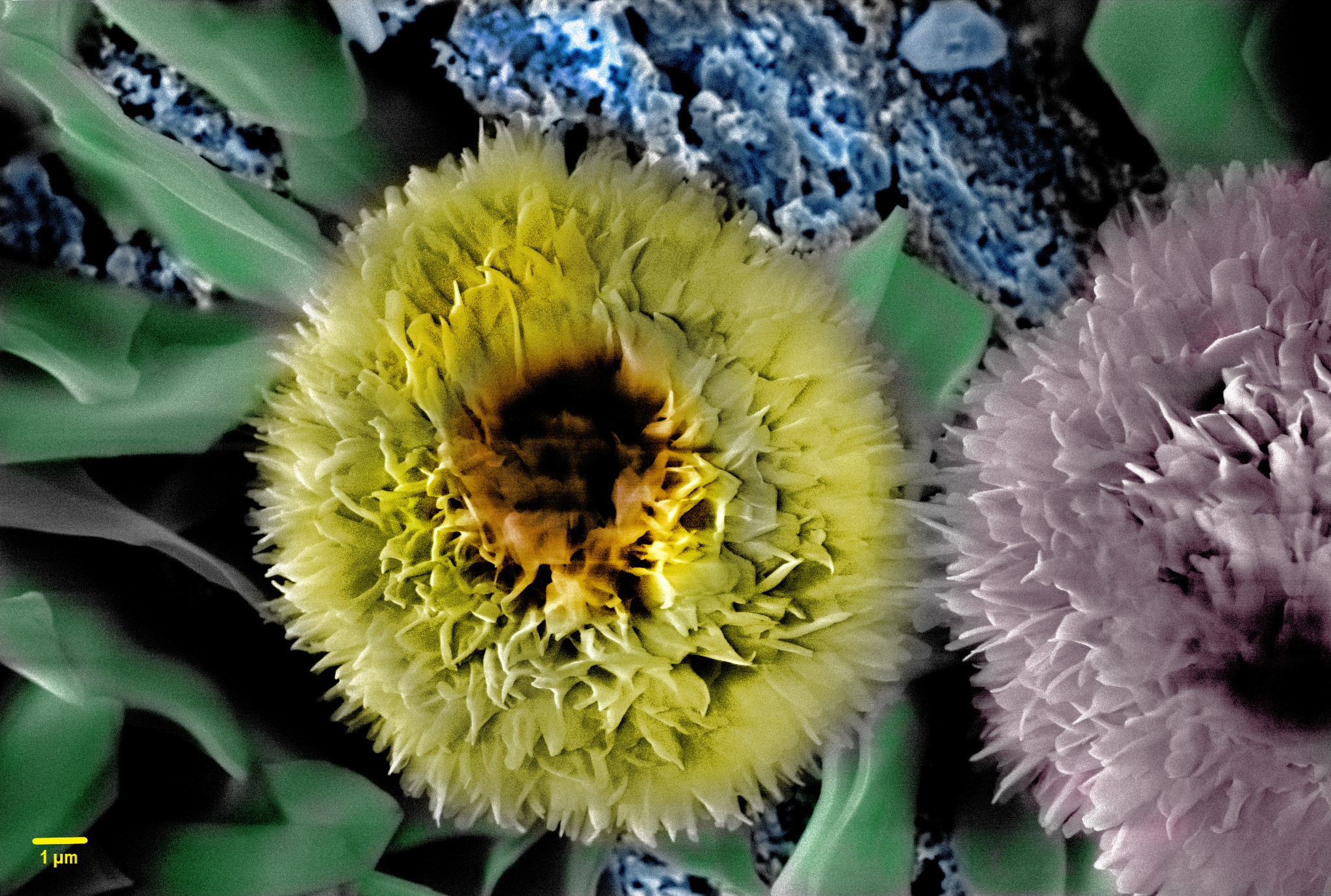
There are Plenty of Sunflowers at the Dark Bottom!
Artists: S.M. Ali Mousavi, Ph.D. Student and Sibin K. Purayil, Post-Doc, Virginia Tech
NNCI Site: NanoEarth
Tool: LEO (Zeiss) 1550 Field Emission Scanning Electron Microscope
One may think sunflowers only grow on soil and lighted earth surface. In the chamber of an SEM and on the pretty dark bottom of brass surfaces, sunflowers were seen growing with nanosized petals all saying “there are plenty of sunflowers at the dark bottom!” to bring hope to all those who need it! The chemisorption of stearic acid on textured etched brass metal surfaces form sunflower-like asperities along nano sheet leaf-like structures.
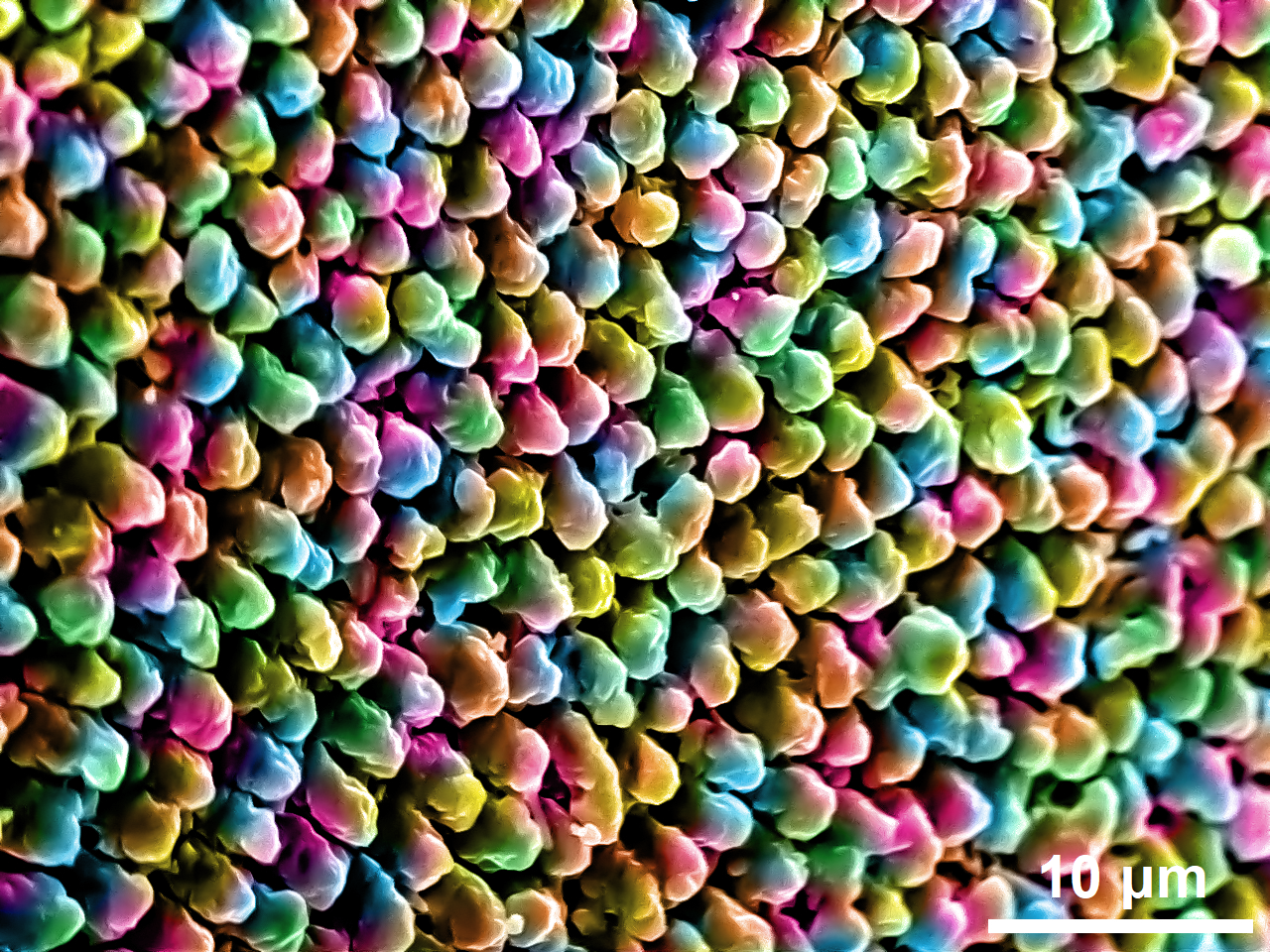
sAlt Water Taffy
Artist: Timothy Lee, PhD Candidate, University of Pennsylvania
NNCI Site: MANTH
Tool: JEOL 7500F HRSEM
Inspired by all the playful colors and flavors of New Jersey salt water taffy, this image makes microstructures look good enough to eat! However, it might taste like metal--all of these lumps are 100% Aluminum (Al). In our lab, we focus on creating metal nanostructures even smaller than the lumps shown in this image that can react fully with water to produce hydrogen gas for various energy applications. Original details were not altered, but rainbow colorization effect was applied.
Gold and Blue
Artist: Matthew Cheng, Student, VPD Research Group, Northwestern University
NNCI Site: SHyNE
Tool: Hitachi SU8030 SEM
Electron microscopy is essential for the observation and analysis of layered van der Waals systems. This is a scanning electron microscope (SEM) image of Cu2Te.
Bouquet
Artist: Erik Boyle, Graduate student, Georgia Institute of Technology
NNCI Site: SENIC
Tool: Zeiss Ultra60 FE-SEM
Nanoelectrospray-assisted focused electron beam-induced deposition is an emerging technique for rapidly growing topologically complex 3D nanostructures from a variety of metallic and nonmetallic materials. A nanoelectrospray delivers the precursor material to the deposition site, where it is irradiated by a focused electron beam, forming a solid phase nanostructure. This image shows a detail near a deposition site of residue left behind by a nanoelectrospray of silver nitrate in a 15% ammonia/85% water solution before post-deposition washing. The drying/cracking pattern in the deposited salt layer and crystalline structures on it evoke budding roses and surrounding leaflets.
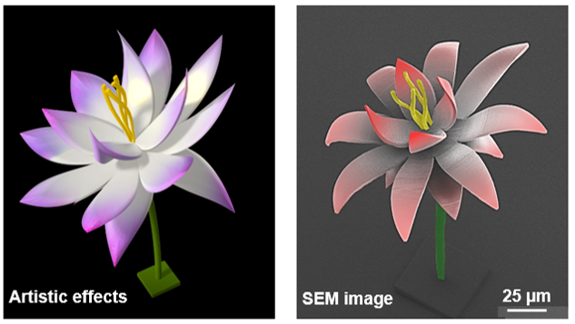
A Micro Blooming Lotus
Artists: Aofei Mao and Peixun Fan, University of Nebraska-Lincoln
NNCI Site: NNF
Tool: Nanoscribe Photonic Professional GT and Hitachi S4700 field emission scanning electron microscope
When engineers become artists, a micro blooming lotus with a dimension smaller than the diameter of human hair was created via a microscale 3D printing technology: two-photon polymerization (Nanoscribe Photonic Professional GT). To make sure the over-hang features of this tiny lotus could survive, Ph.D. student Aofei Mao, Dr. Peixun Fan, and Prof. Yongfeng Lu from the Laser Assisted Nano-Engineering Lab invented their “gradient polymerization” technique by finely controlling the printing parameters. The sample was sputtering-coated for SEM imaging using a Hitachi S4700 field emission scanning electron microscope.
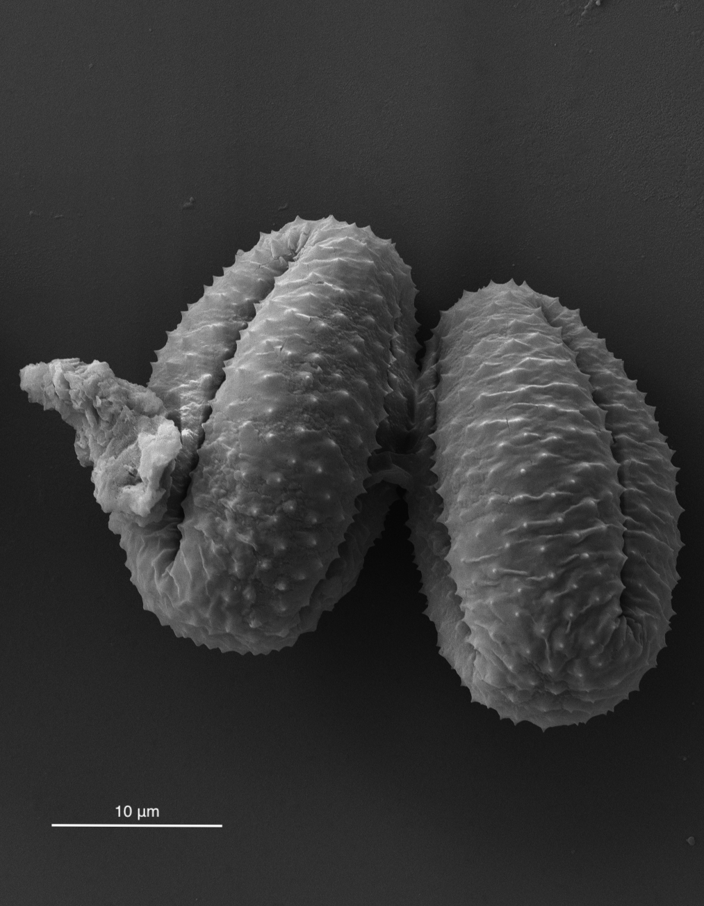
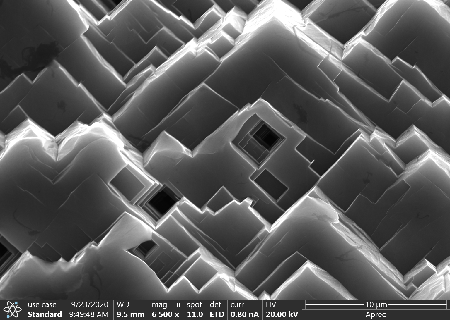
Lungs of Nature
Artists: Hanna Varga, student, Duke University
NNCI Site: RTNN
Tool: Apreo S by ThermoFisher Scientific Scanning Electron Microscope
The image depicts particles from a dust sample collected on the California coast. This pair of pollen grains reminded the researcher of lungs.
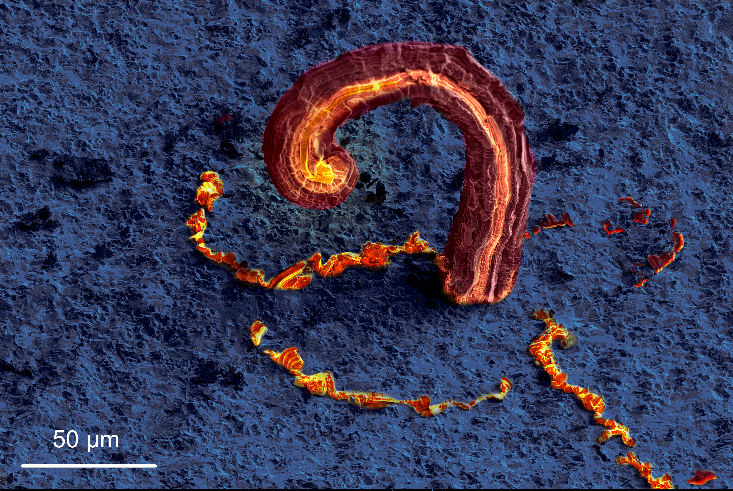
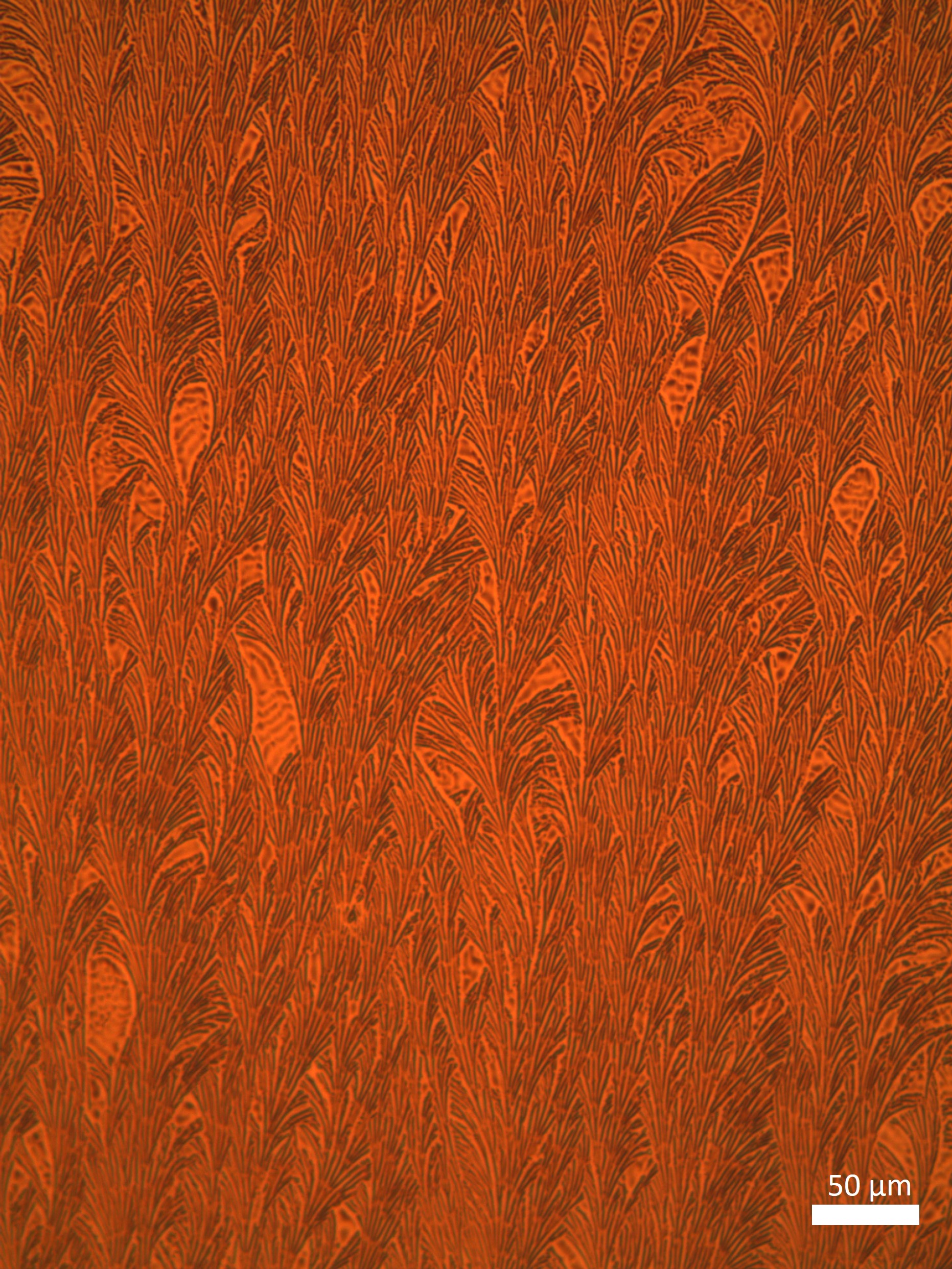
Lithium Nightmares
Artist(s): Xin Xu, Postdoctoral Fellow, and Geoff McConohy, Graduate Student, Materials Science and Engineering, William Chueh Group, Stanford University
NNCI Site: nano@stanford
Tool: FEI Helios NanoLab 600i DualBeam SEM/FIB
This image shows the promise and peril of a new kind of lithium battery using a ceramic solid electrolyte. The dark region in our image is the electrolyte while the orange structures are lithium metal, which grows up from the electrolyte by applying an electric voltage. First, the large lithium metal whisker grows rapidly, which is good for a fast-charging battery. But at some point, the lithium wire stops growing and instead the lithium is seen cracking the electrolyte as it grows through the ceramic: the spooky filaments are destined to destroy the battery.
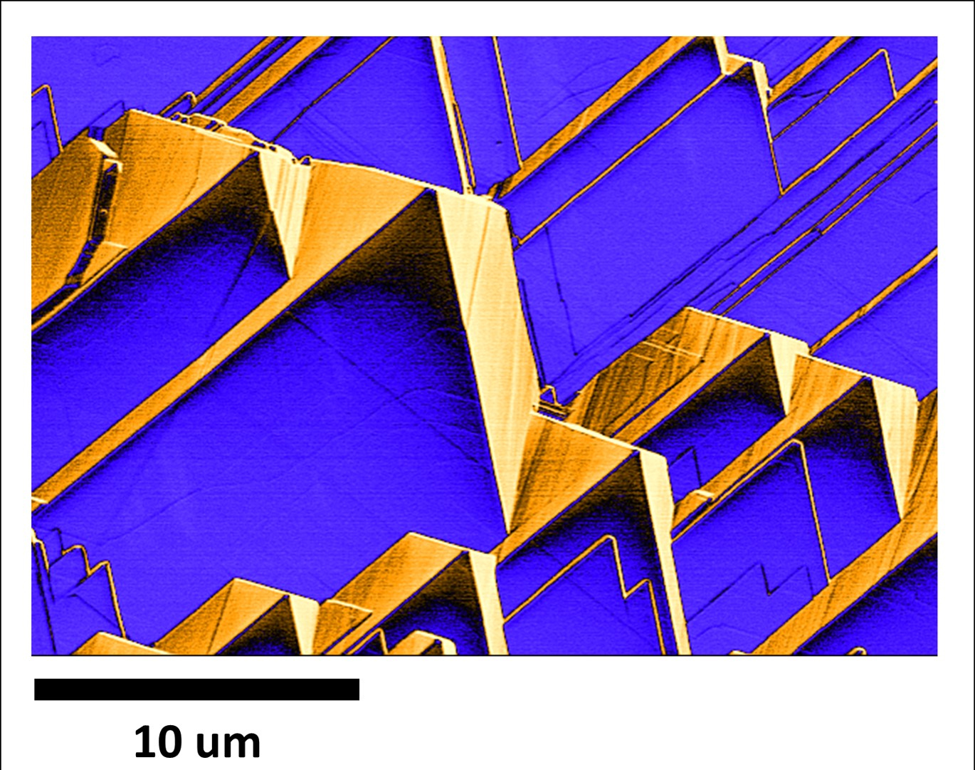
Nano Adobies
Artist: Mike Martin, Huson Lab, University of Louisville
NNCI Site: KY Multiscale
Tool: Apreo SEM
These structures were found on an aluminum sputtering target after etching in aqua regia.
Dendrites168
Artists: Brian Torrini, Undergrad research assistant, Holmes Research Group Lab, University of Minnesota
NNCI Site: MiNIC
Tool: Optical Microscope
This was taken in Professor Holmes' lab under an optical microscope. A thin film of an electron transport material known as TPBI, used in optoelectronics, was deposited onto a Si substrate and annealed. Pictured is about 30 nm of TPBI annealed to 168 ℃. Professor Holmes’ research group was interested in a regular repeating pattern formed during the annealing process that resembles ripples on a pond. What is pictured here is a dendritic phase, which was not an area of interest, but the patterns and color made an intriguing picture.
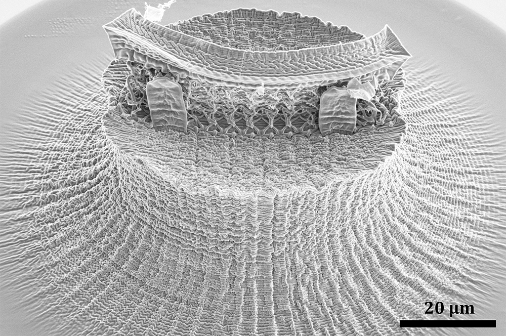
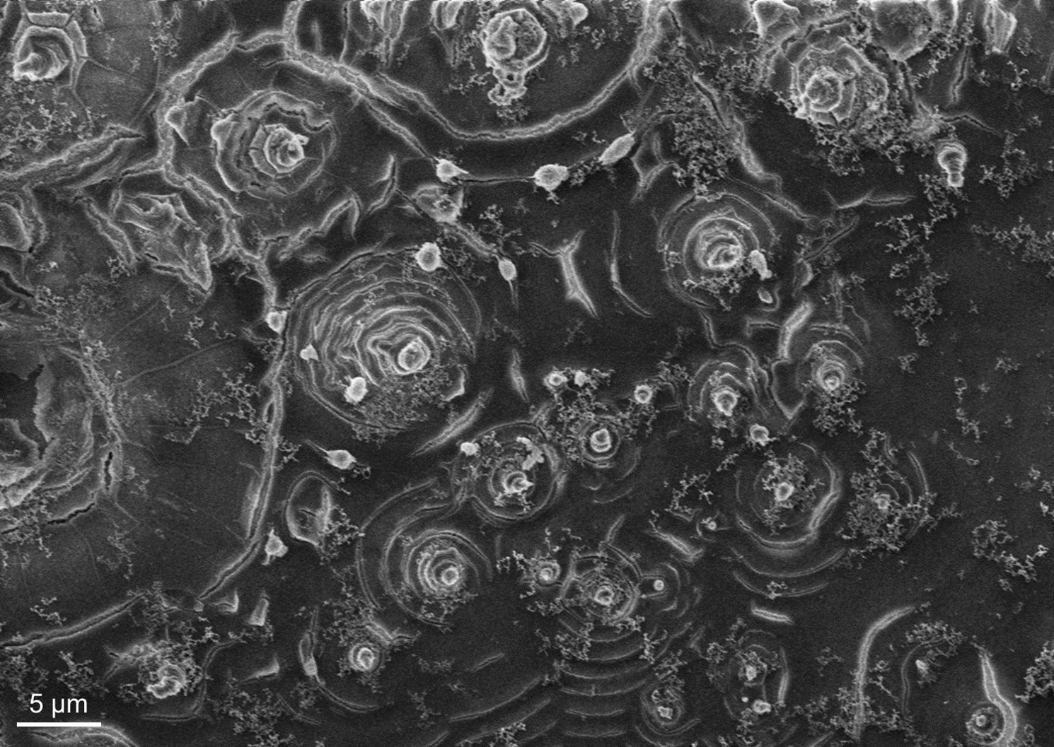
Nano-wrinkled Head
Artists: Zainab Patel, Graduate student, and Lucas Meza, University of Washington
NNCI Site: NNI
Tool: Apreo SEM
A 3D printed fracture test assembly was plasma etched which formed a thick skin on the printed polymer. When this assembly was pyrolyzed at 900C to shrink the polymer, the skin buckled and formed this unique and highly textured surface never seen before, which seems like a wrinkled human head.