
NNCI Image Contest 2020 - Unique
Most Unique Capability
This category celebrates the unique capabilities each site of the NNCI has on offer. Please check out the images below and read a little about the research behind them.
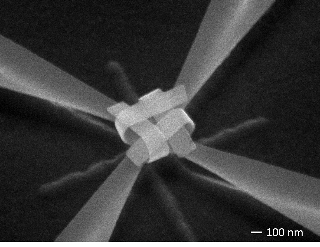
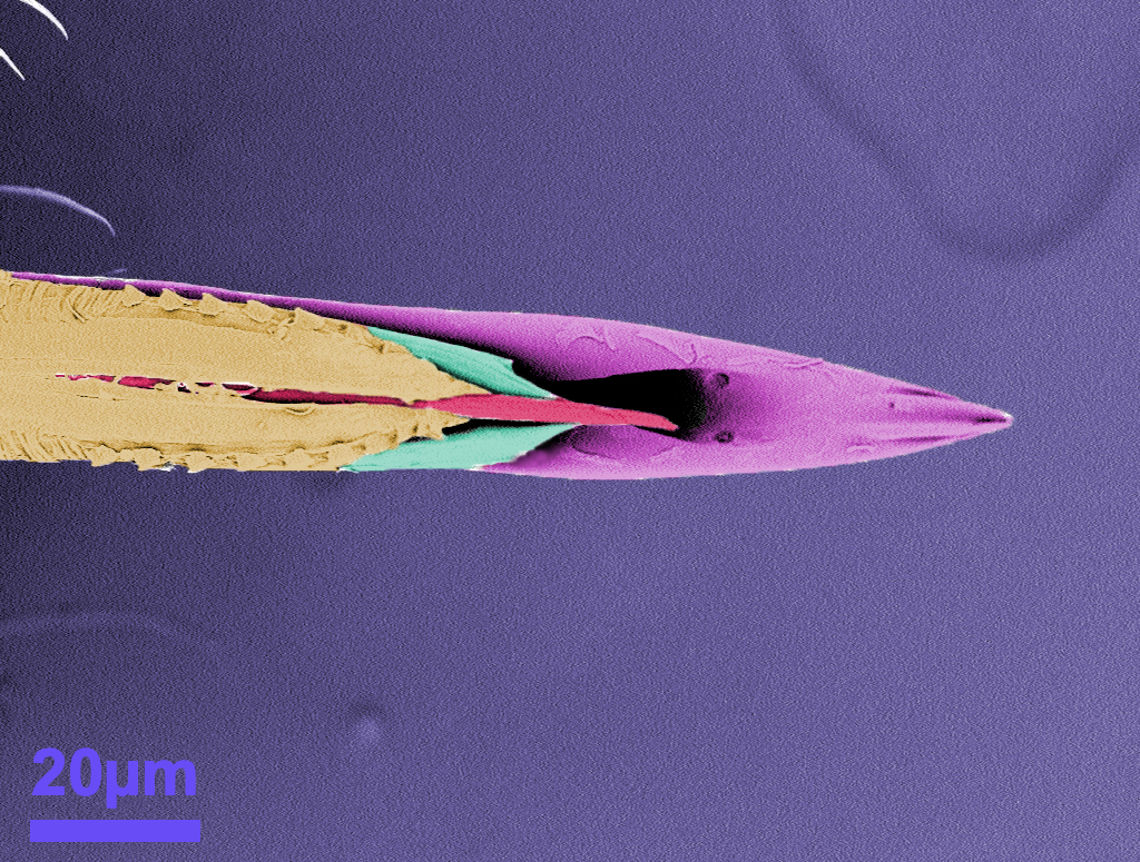
Electron Irradiation Driven Hands for Nanoweaving
Artists: Chunhui Dai, graduate student, and Jeong-Hyun Cho, faculty member, Department of Electrical and Computer Engineering, University of Minnesota, Twin Cities
NNCI Site: MiNIC (University of Minnesota Characterization Facility)
Tool: FEI Helios Nanolab G4 dual-beam focused ion beam (FIB)
Inspired by the idea to scale down factories by cascading manipulator hands with diminishing scale, suggested by Nobel Laureate Dr. Feynman, we developed a method that uses electron irradiation as “hands” to manipulate nanostructures. In this image, a nanoknot has been woven using an electron beam. The mechanism is based on irradiation-induced crystallization of amorphous aluminum oxide (Al2O3) in released bilayers (chromium, Cr/Al2O3), which generates localized stress for self-assembly. This work brings nanoscale origami-like self-assembly to a new level, which could trigger innovations in the fields of nanomachines, nanoscale test platform, and advanced optical devices.
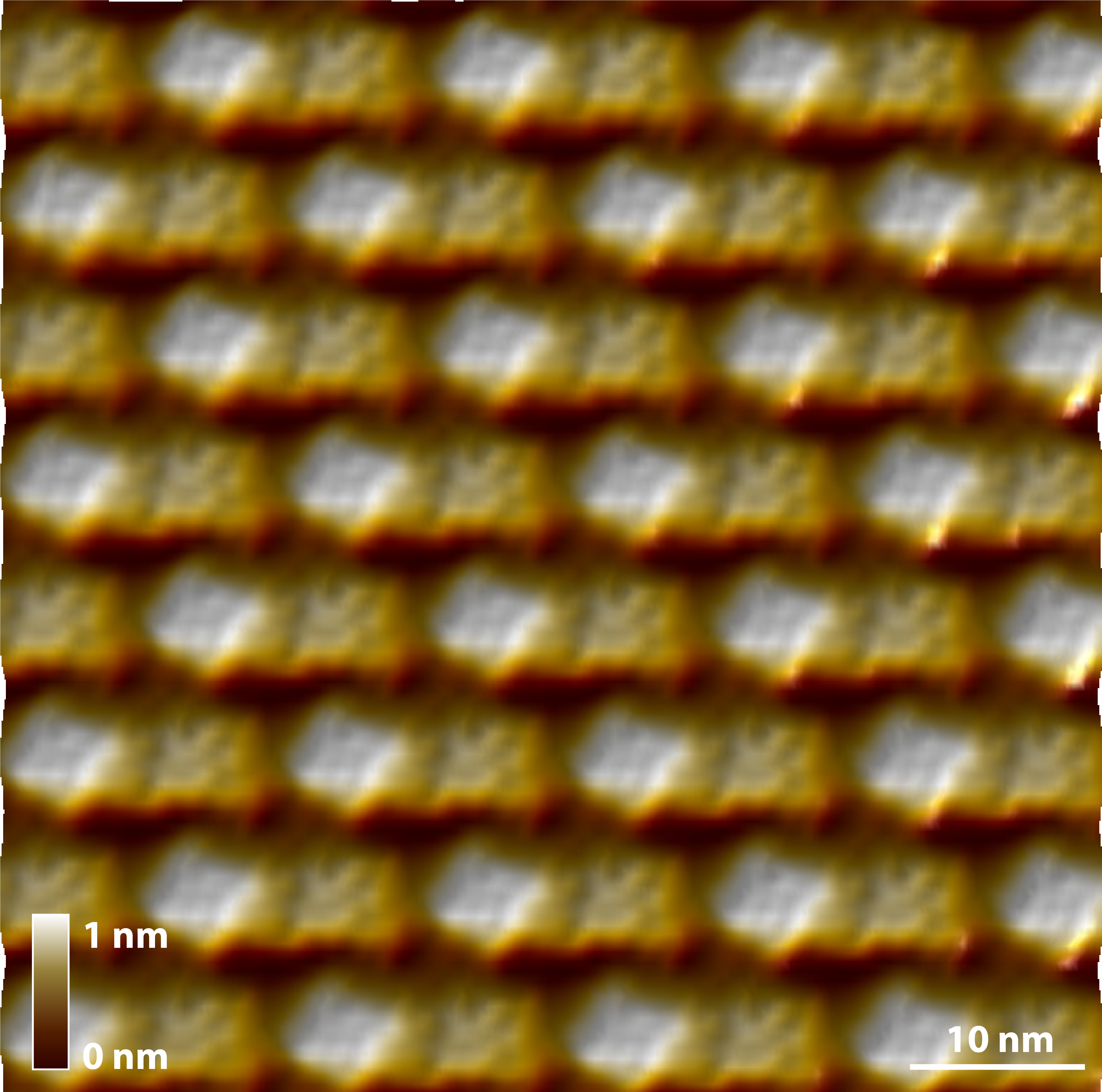
“GLAD” to help Mother Nature do Nanoscience!
Artist: Dr. Chuang Qu, University of Louisville
NNCI Site: KY Multiscale MNTC Cleanroom and Huson Imaging and Characterization Lab
Tool: Kurt Lesker e-beam Evaporator with Angle Control and Thermo Fisher Apreo C Low Vac SEM
Glancing Angle Deposition (GLAD) of germanium on line seeds demonstrating how a variety of self-assembled micro and nano structures can be created by adjusting the seed morphology, material and angle of deposition.
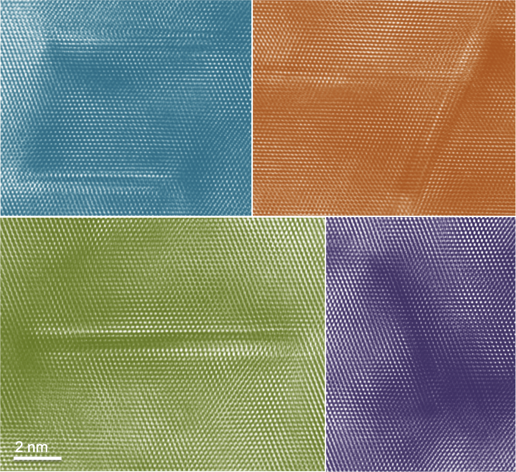
Desirable Atomic Defects
Artists: Pariasadat Musavigharavi, Postdoc, University of Pennsylvania
NNCI Site: MANTH
Tool: JEOL F200 TEM
The image entitled “Desirable Atomic Defects” demonstrates a network of planar defects in Aluminum thin film. A perfect crystal, with every atom of the same type in the correct position, does not exist. Defects are observed in all crystal structure. Defects are used in ways that are vital for device fabrication. Transmission Electron Microscopy is employed for studying the defects in different scales. In the previous studies, researchers used to decline or eliminate defects that can degrade properties, although this approach is turned on its head; creating materials with “designer defects” that are the functional portion of the device.
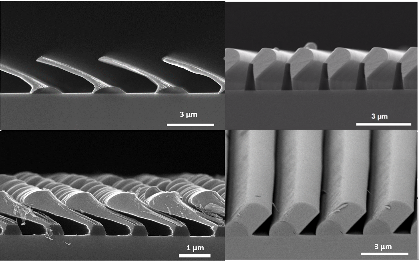
Standing Tall
Artist: Austin Hickman, Graduate Student, Cornell University
NNCI Site: Cornell NanoScale Facility (CNF)
Tool: Zeiss Ultra SEM
The T-gate, named for the rough outline of its cross-section, is the fundamental device component of high-frequency transistors. The T-gate pictured here, composed of titanium and gold, shows a large, 200x250 nanometers (nm) "head" sitting atop an astonishingly thin "stem", just 22 nm in length and 125 nm tall. The most significant measurement is the gate length, which rivals that of the shortest T-gate length ever demonstrated by industry or academia, 20 nm. This short gate length will enable high-frequency transistor performance with a target application of the sixth-generation (6G) wireless network.
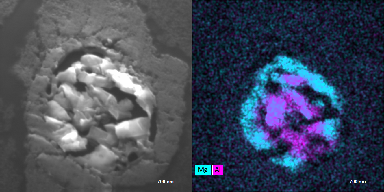
We are All Star Dust
Artist(s): Nathaniel Rieders, PhD candidate, and Recep Avci, PI at Montana State University
NNCI Site: MONT
Tool: Integrated Auger Nanoprob
Interplanetary dust particle from the pre-solar nebula. This particle predates our own solar system and was collected in the stratosphere by U2 spy planes. EDX maps show heterogeneous distribution of Al and Mg. Sample courtesy of Dr. David Joswiak (University of Washington).
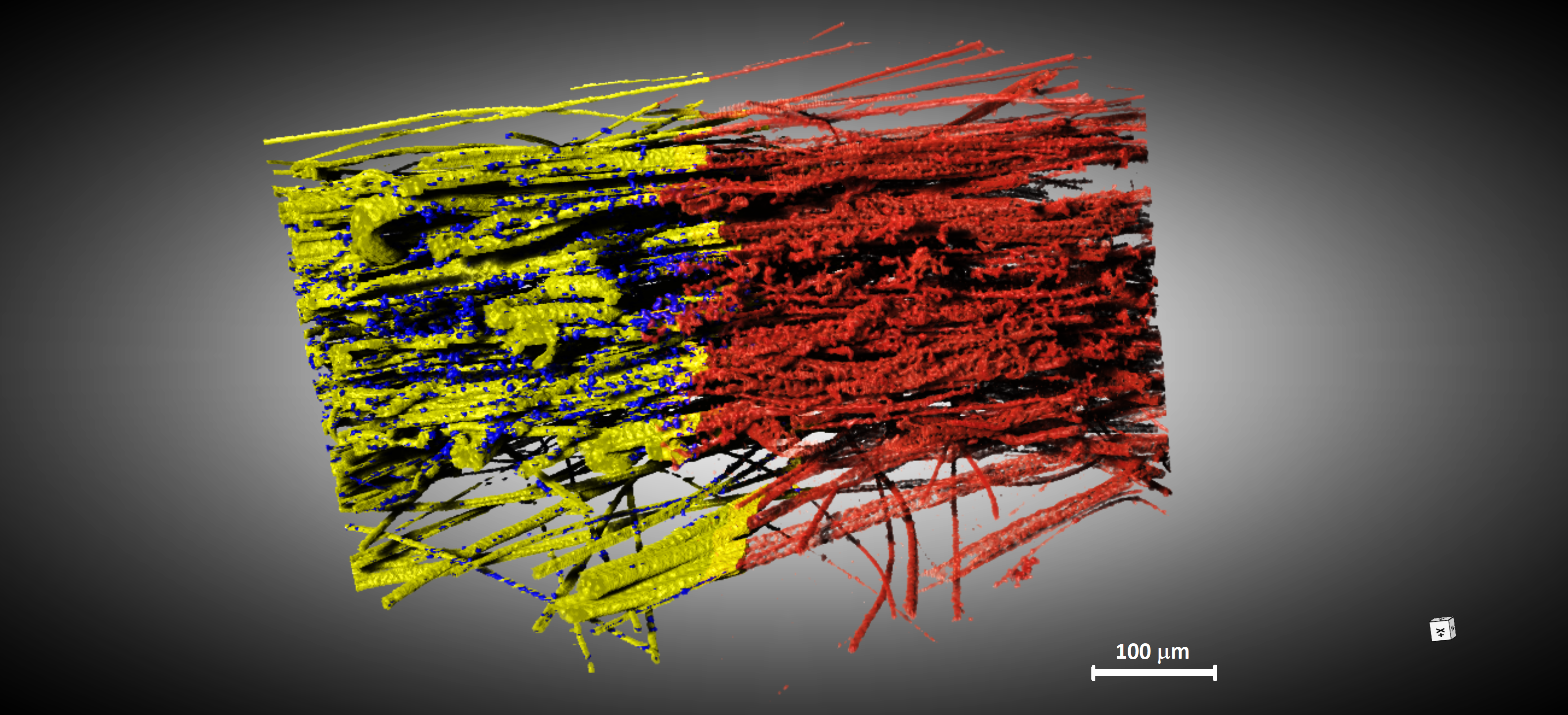
Revealed Inner Secret (Segmentation and classification)
Artist(s): Hye Ryoung Lee, Scientist, Stanford University and Arturas Vailionis, Senior Research Scientist, Stanford Nano Shared Facilities
NNCI Site: nano@stanford
Tool: Zeiss Xradia 520
Filtering facepiece respirators (FFR) became our daily life items in the COVID-19 pandemic era. We analyzed a meltblown filter fabric layer in N95 respirators by X-ray microscope. For additional details, we used deep learning (DL) algorithms to distinguish particles from fibers. Red color indicates the reconstructed 3D-X ray image which contains both fibers and particles. Blue color shows DL-assisted segmented NaCl particles which were used as contaminants for filtration efficiency test and yellow color is DL-assisted segmented polypropylene fibers.
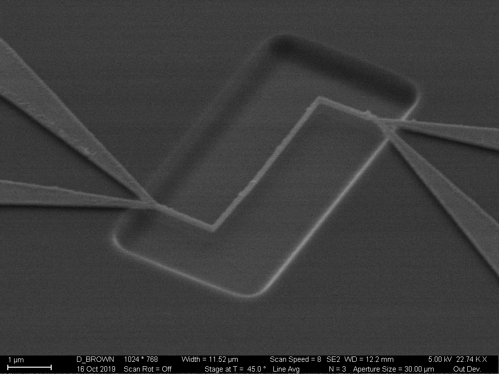
Suspended Nickel Nanowires on Oxide Substrate
Artist(s): Devin K. Brown, Ph.D. Electrical and Computer Engineering student, Georgia Institute of Technology
NNCI Site: SENIC
Tool: Imaging tool: Zeiss Ultra60 FE-SEM, Key patterning tool: Elionix ELS-G100 100 kV electron beam lithography system
The image shows a nickel wire 100 nm in width, suspended over an etched cavity in a thermally grown 500 nm silicon dioxide film. The nickel nanowire is designed to serve as a piezo-resistive force sensor for blood platelet compressive force transduction. This information would be useful for blood disease diagnosis. The nanowire pattern was created using an Elionix ELS-G100 100 kV electron beam lithography system in combination with a liftoff process. The nanowire metal consists of an adhesion layer of 40 Å of titanium followed by 1000 Å of nickel deposited by a CVC electron beam evaporator.
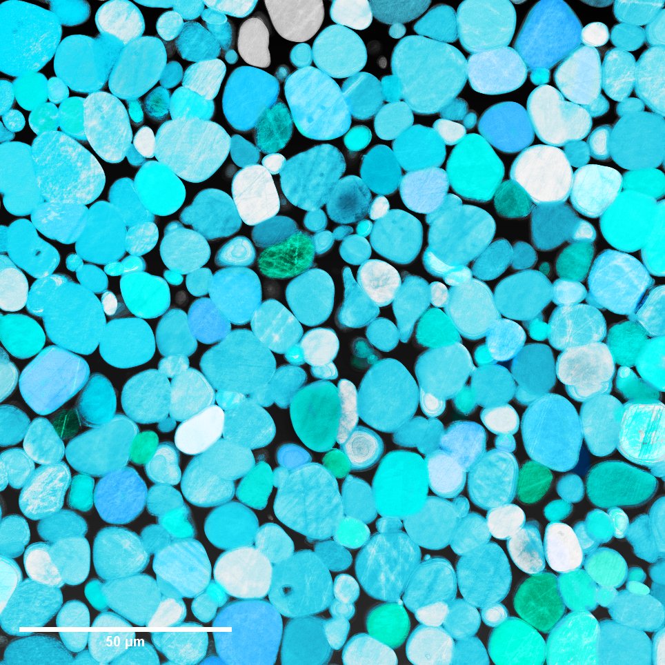
River Rocks, or Nuclear Grade Alloy?
Artist: Jacob Haag, Ph.D. student, Materials Science Engineering, Virginia Polytechnic Institute and State University
NNCI Site: NanoEarth
Tool: FEI Quanta 600 FEG Environmental SEM
If you guessed nuclear grade alloy, you're right! This is an image depicting a 3D reconstruction of a specialized composite called a tungsten heavy alloy. The semi-spherical domains are single grains of tungsten, which have been suspended in a sponge-like network of nickel and iron. This structure gives these alloys the incredible ability to be both hard and ductile at the same time, making them perfect choices for demanding environments. These materials showcase nanoscale beauty and complexity along with amazing functionality!
Elegant Mosquito Fascicle
Artist: Kun Luan, Post-doc, NC State University
NNCI Site: RTNN
Tool: Hitachi TM4000 Tabletop SEM
"Elegant Mosquito Fascicle" reveals the micro-anatomy of mosquito stylet. It can explain how the mosquito bites through human skin by using proboscis. The information conveyed from the image were used to engineer non-insecticide barriers, which can mechanically prevent the mosquito bite.
Protein Chess Board in Nanoscale
Artist: Shuai Zhang, Faculty Member, University of Washington
NNCI Site: NNI Molecular Analysis Facility
Tool: Cypher atomic force microscopy
An atomic force microscopy (AFM) averaged morphological image of the two-dimensional protein crystals of mutated l-rhamnulose-1-phosphate aldolase (F88/C98RhuA) on muscovite mica. It was captured by Cypher AFM in the buffer of 50 mM MgCl2 and 20 mM Tris (pH 7). The sample was provided by the Tezcan lab at UCSD.
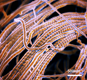
Decellularized Mouse Spinal Cord Tissue
Artist(s): Zaida Alvarez Pinto, PhD, Research Assistant Professor, Northwestern University, Simpson Querrey Institute & Alexandra Kolberg-Edelbrock, PhD, Former Graduate Student, Northwestern University, Simpson Querrey Institute
NNCI Site: SHyNE
Tool: Hitachi SU8030
This image shows a close-up of mouse spinal cord decellularized extracellular matrix. The bundled fibers with regular banding features make up the extracellular matrix in the spinal cord and function to support cells such as neurons and glial cells within this tissue. Scientists can use this image to design new materials that mimic the natural architecture of spinal cord tissue. These materials can be used in the context of developing new regenerative medicine therapies to treat spinal cord injury.
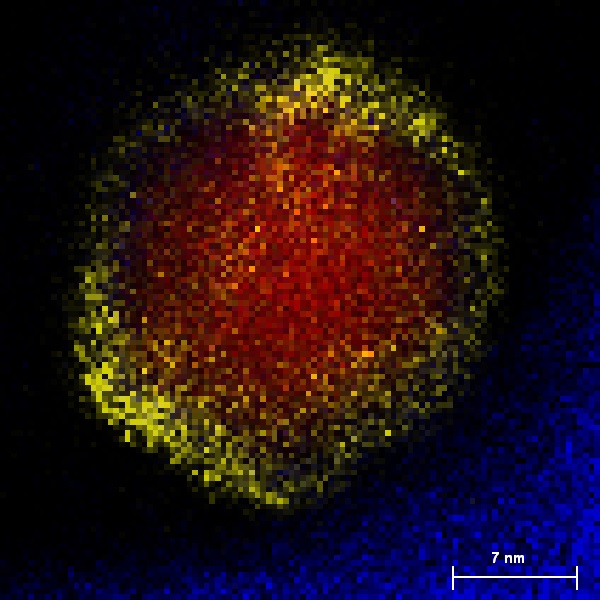
The Sunset
Artist: Zahra Ahmadi, Graduate student), University of Nebraska-Lincoln
NNCI Site: Nebraska Nanoscale Facility
Tool: FEI Tecnai Osiris (Scanning) Transmission Electron Microscope
The image shows energy dispersive X-ray spectroscopy (EDS) elemental mapping of a core/shell Co/ZnO nanoparticle created using FEI Tecnai Osiris (Scanning) Transmission Electron Microscope. The nanoparticle was formed in an inert-gas condensation chamber located in the material processing lab in department of mechanical and materials engineering at UNL. The core of the nanoparticle is made up of Co which is shown in red. ZnO shell surrounded the core, however only the Zn is shown in this image with yellow color. The blue color at the bottom represents the oxygen.