
NNCI Image Contest 2019 - Stunning
Most Stunning
This category celebrates the beauty of the micro and nanoscale. Click on the most stunning image in order to vote for it. You may vote for only one image in this category.
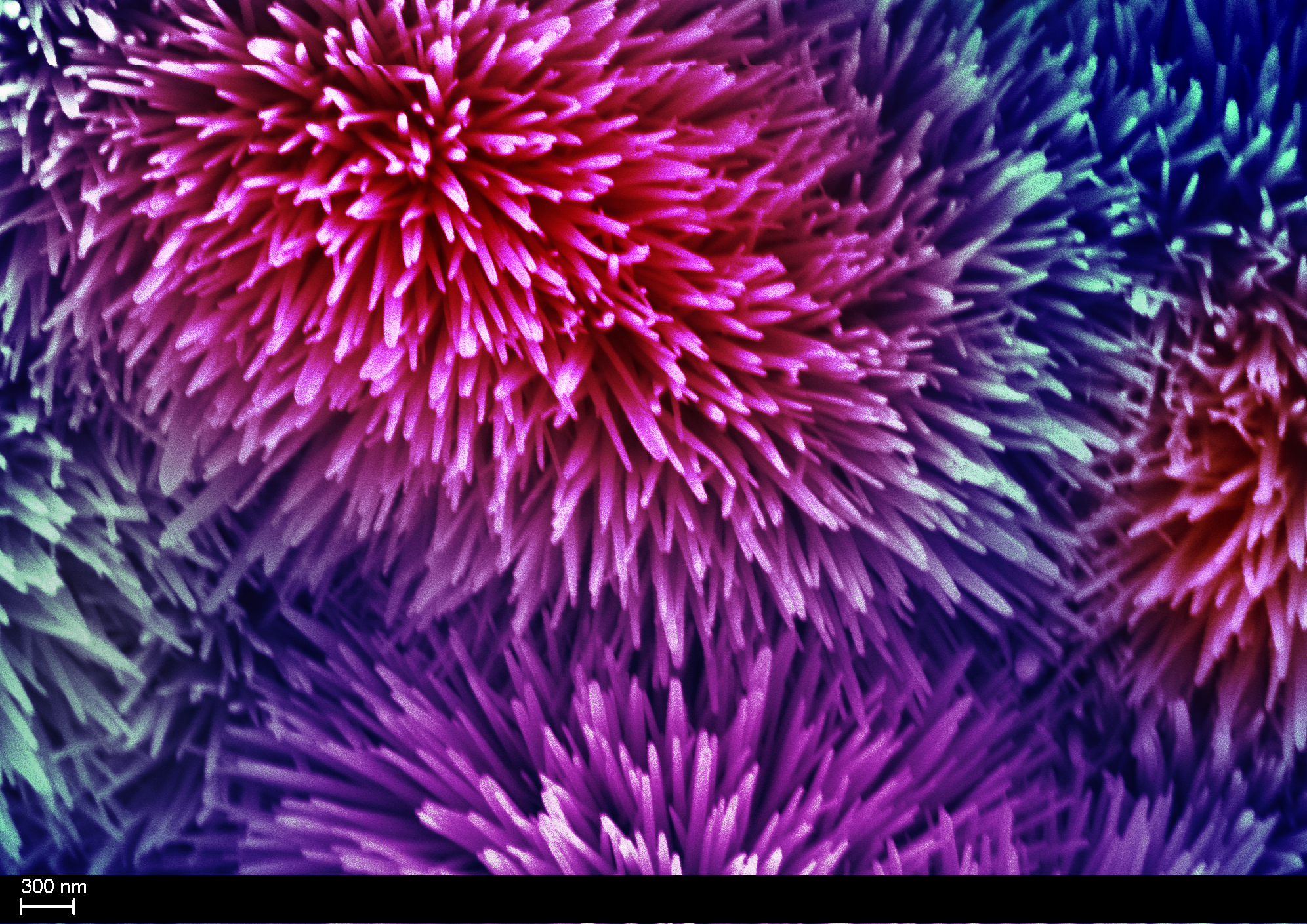
Nano Fireworks
Artist: Yamin Zhang, PhD Student in School of Chemical and Biomolecular Engineering, Georgia Tech
NNCI Site: SENIC
Tool: Zeiss Ultra 60 FE-SEM
This image consists of many rods. The diameter of the rod is around 100 nanometers. So, they are called nanorods. The material of these nanorods is zinc oxide. Zinc oxide nanorods grow on a flat nickel metal substrate naturally and nicely, which look like fireworks. In our research, zinc oxide nanorods are used as the anode material for zinc-based aqueous batteries, which are a safe alternative for current lithium ion batteries. With well aligned structure, the morphology change of zinc oxide nanorods anode during battery cycling can be easily studied by imaging it, which explains capacity loss of batteries.
The Copper Garden
Artist: Loic Constantin, Graduate Student in the Lane Lab, Univ. of Nebraska-Lincoln
NNCI Site: NNF
Tool: FEI Quanta 200 Environmental SEM
Copper particles melted on titanium coated carbon fibers. The demand to develop new materials for thermal management is crucial to nuclear, microelectronic and aerospace applications. Creating a composite material consisting of two highly thermal conductive elements, such as copper (Cu) and carbon (C), may fulfill these unmet needs. However, the lack of chemical affinity between Cu and C presents a major challenge. To resolve this issue, we coated carbon fibers (CFs) with titanium via an innovative molten salt process. The titanium layer creates chemical bonds between the Cu and CFs, which facilitates heat transfer in the final composite materials.
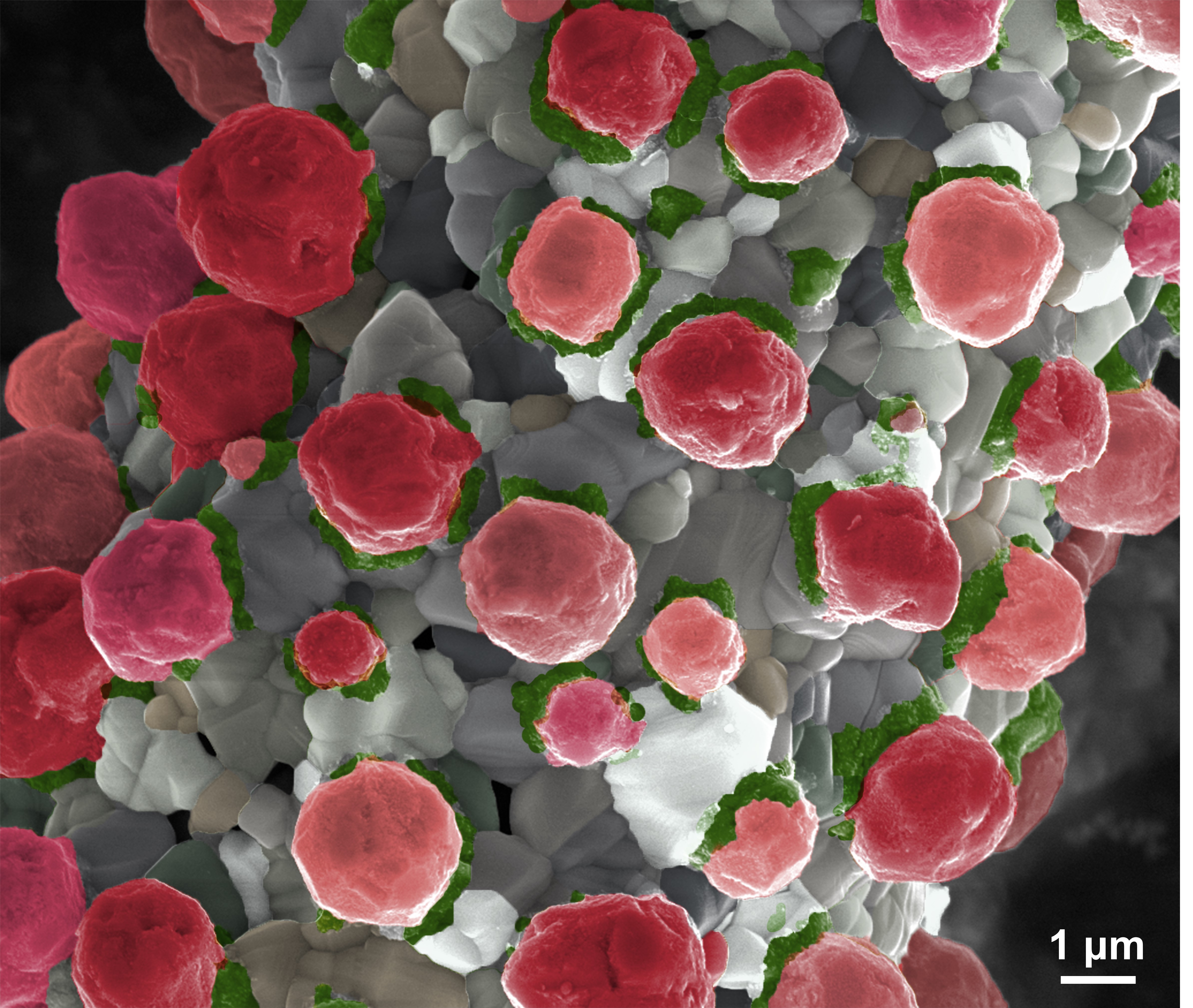
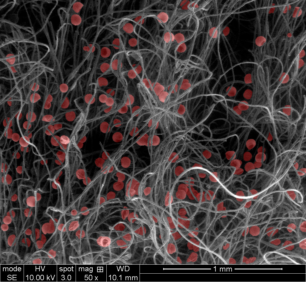
Cyborg Microorganisms: Use of Rotifers as Self-Propelling Biohybrid Microcleaners
Artists: Fernando Soto, Graduate Student, and Ryan Nicholl, Nano3 Staff, University of California-San Diego
NNCI Site: SDNI
Tool: FEI Quanta FEG 250 Scanning Electron Microscope
Taking inspiration from the ‘Sci-fi’ concept of a cyborg, where an organism has enhanced abilities due to the integration of some artificial component. We developed a self-propelled biohybrid microrobot by combining the mobile marine rotifers (blue) with functionalized microbeads (yellow), turning the Cyborg into a micro Roomba (cleaning robot)!
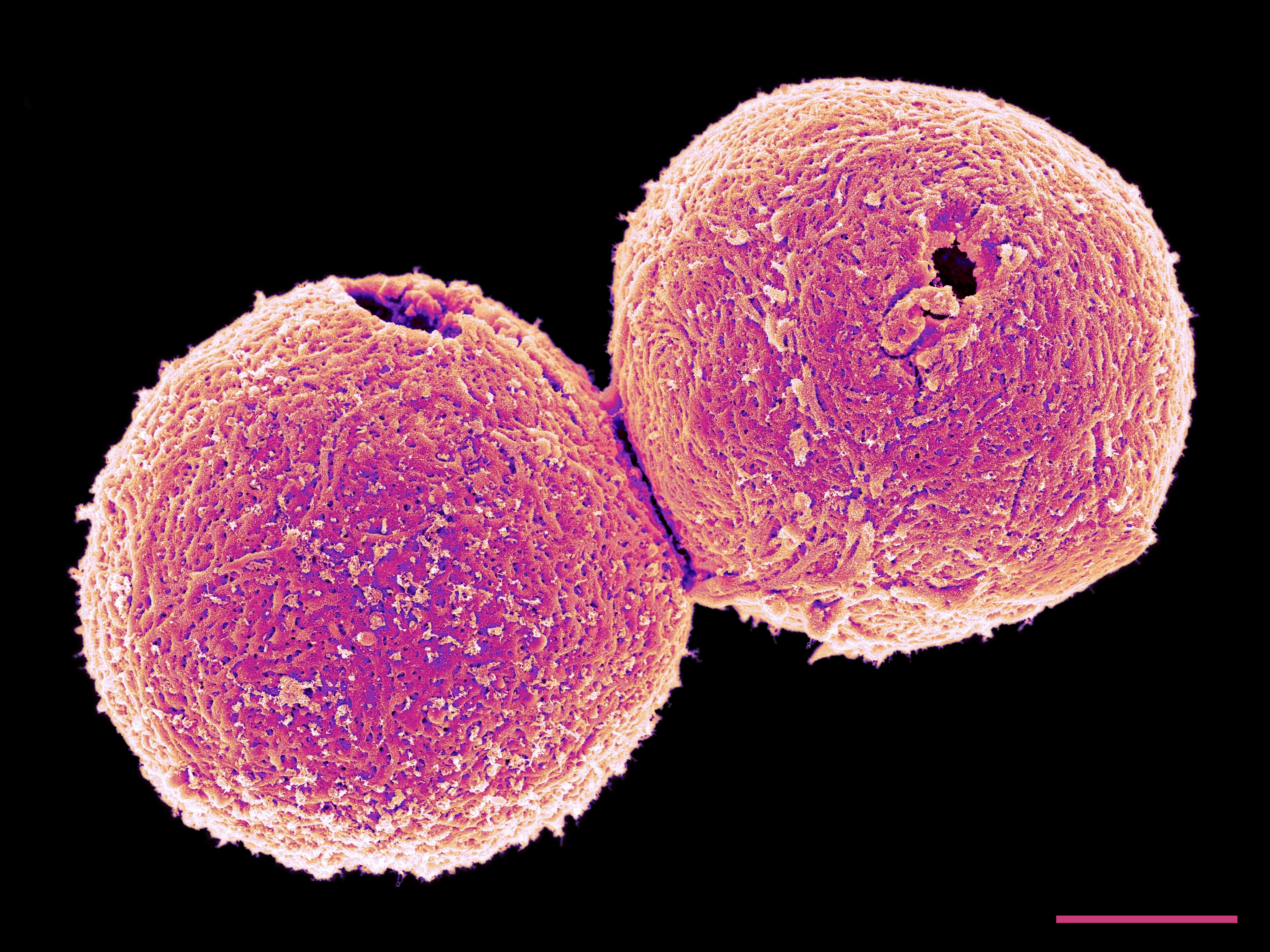
A "Hole" New Method for Sperm Storage
Artist: Eric W. Roth, BioCryo Electron Microscopy Specialist, Northwestern University
NNCI Site: SHyNE
Tool: HITACHI SU8030 SEM
Cancer treatments, while life preserving, can threaten fertility. Fertility preservation can be challenging in males with conditions of extremely low germ cell numbers or in those who have undergone testicular or epididymal biopsy where there may be limited numbers of immature germ cells. A significant clinical hurdle is the storage and recovery of small numbers of sperm. We engineered an oocyte-derived biomaterial – the zona pellucida (ZP) – to function as a “sperm safe” for storing sperm. The ZP is a glycoprotein matrix that surrounds the mammalian oocyte. Using a piezo drill, we made a small hole in the ZP and mechanically separated it from the oocyte cytoplasm and then further removed cellular material using a decellularization process. This images shows two deceullarized sperm safes imaged by scanning electron microscopy. The holes in which sperm are deposited within the “sperm safes” are clearly visible. Scale bar equals 10um.
Gill Raker of the Japanese Medaka
Artist: Melissa Chernick, Research Technician, Duke University
NNCI Site: RTNN
Tool: FEI XL30 SEM-FEG
This image shows a portion of a gill raker from a Japanese medaka (Oryzias latipes), a small fish often used as a research model. Gill rakers are tooth-like structures inside a fish that help capture prey and prevent damage to its gills. The image shows some of the tissue that makes up the gill raker: a taste bud surrounded by pavement cells.
Therapeutic Fruits
Artists: Yun Kee Jo, Postdoc, and Daeyeon Lee, faculty member, Chemical and Biomolecular Engineering, University of Pennsylvania
NNCI Site: MANTH
Tool: Quanta 600 FEG ESEM
Therapeutic drug-loaded microcapsules are entrapped in a biomedical gauze patch. They would release the therapeutics cleverly upon mechanical forces from the movements of the gauze such as stretching or bending.
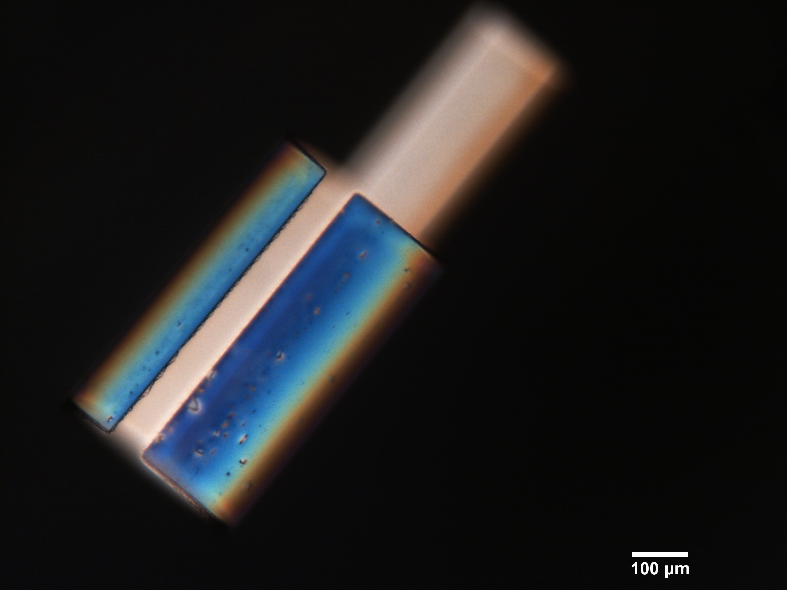
HugBot
Artist: Anna Alvarez, CNF REU, University of Illinois at Urbana-Champaign
NNCI Site: CNF
Tool: Olympus MX-50
This image depicts "Hug-Bot", a thermal responsive liquid crystal film that was developed and photo-patterned at CNF. Exposure to heat induces a phase transition in the liquid crystal bimorph, causing the film to buckle and curve along a predetermined alignment direction. Hug-Bot is the first successful test of these new responsive films and serves as a stepping stone for future structures patterned with this material. The image was taken via the Olympus MX-50 in the polarizing optical microscopy mode.
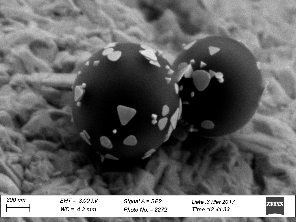
Ultra Low Calorie Nano Cake Pops
Artist: Emtias Chowdhury, graduate student, University of Louisville
NNCI Site: KY Multiscale
Tool: Zeiss Supra 35 scanning Electron Microscopy
Anisotropic Nanoprism Coated Polymer Beads. DNA mediated interaction between nanoscale gold nanoprisms and microscale polymer beads.
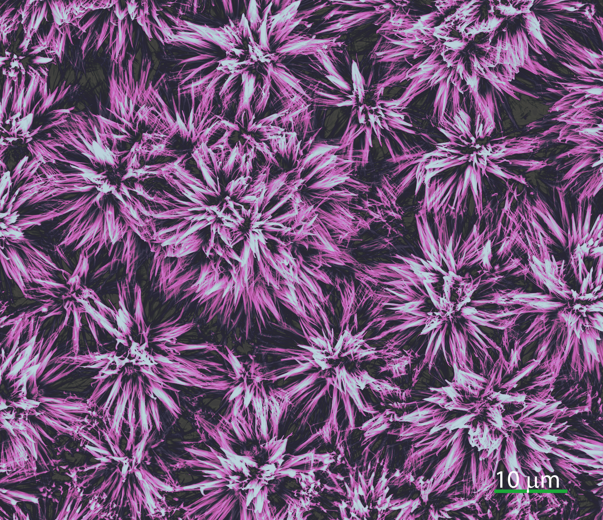
Dahlia
Artist: S.M.Ali Mousavi and Jonathan Angle, Materials Science & Engineering Graduate Students, Virginia Tech
NNCI Site: NanoEarth
Tool: FEI ESEM
A scientist engineer also likes to take pictures of mother nature's articraft that is built in the lab. SEM images of long chain hydrocarbons (from stearic acid solution) chemisorbs on a bed of cauliflower and grows in the shape of a Dahlia flower that mimics the superhydrophbicity of Lotus (Nelumbo nucifera) leaf are shown.
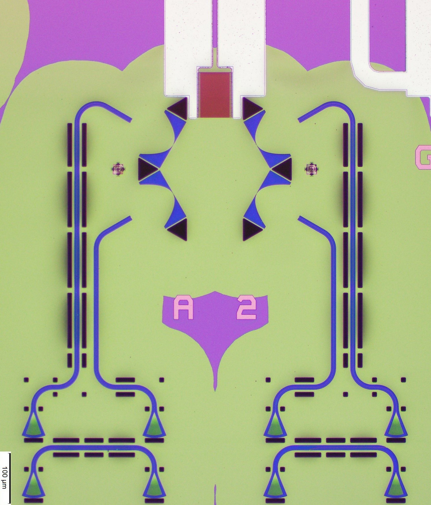
An Acousto-Optic “Flower”
Artist: Huan Li, Research Associate in the Department of Electrical and Computer Engineering, and Mo Li, Associate Professor in the Department of Electrical and Computer Engineering and Department of Physics, University of Washington
NNCI Site: NNI
Tool: Olympus optical microscope onsite demonstration at the Washington Nanofabrication Facility
This is a true-color image of a new-generation integrated acousto-optic prototype device designed to meet crucial sensing and communication challenges. The “flower” is fabricated on a 330 nm thick suspended piezoelectric membrane, which is the large green area within the purple background. From the four small blue petals and the large red and white petal, respectively, light and sound waves of carefully chosen wavelengths are injected into the center of the flower, where light propagation is bent by sound of ultrahigh frequency by 60°, manifesting an intriguing physical phenomenon called Brillouin scattering in an unprecedented regime.
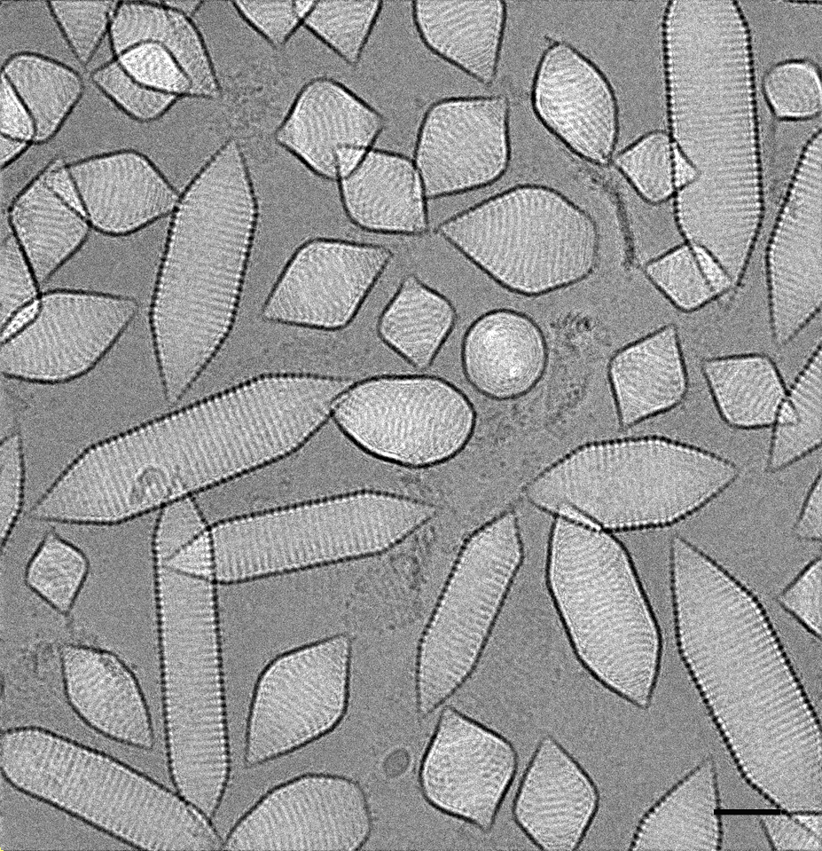
The (Gas Vesicle) Lilies in homage to Claude Monet
Artist: Guanhong Bo, PhD student, School for Engineering of Matter, Transport and Energy, Arizona State University; Brent Nannenga, Asst. Professor, School for Engineering of Matter, Transport and Energy, Arizona State University; and Dewight Williams, Research Scientist, Eyring Materials Center, Arizona State University
NNCI Site: NCI-SW
Tool: Titan Krios cyrogenic TEM at Arizona State University
Images of gas vesicles from bacteria imaged by cryogenic electron microscopy. Gas vesicles are hollow gas filled tubes made of protein that certain bacteria use to regulate their buoyancy. Samples were provided by Mikhail Shapiro (California Institute of Technology). The scale bar is 40 nm.
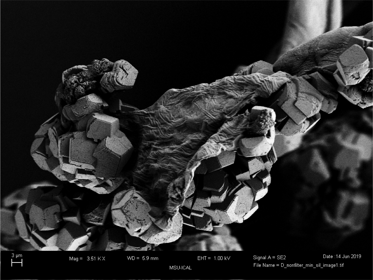
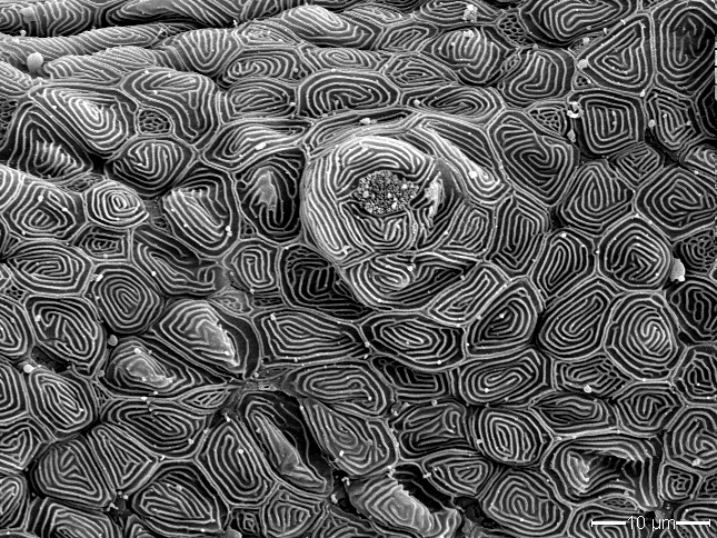
Mineralized Architecture
Artist: Chelsea Heveran, PhD, and Michael Espinal, undergraduate, Montana State University
NNCI Site: MONT
Tool: Zeiss Supra 55VP
Can biomineralized agricultural waste be used to reinforce cement? Here, we biomineralized cellulose fibers using jackbean enzyme, and then coated the mineralized fibers with a thin hydrophobic silane film. These biomineralized fibers are inspired by the mineralized fiber architecture of bone and may lead to hierarchical toughening of cement. Image acquired using a Zeiss Supra 55VP at MSU ICAL.
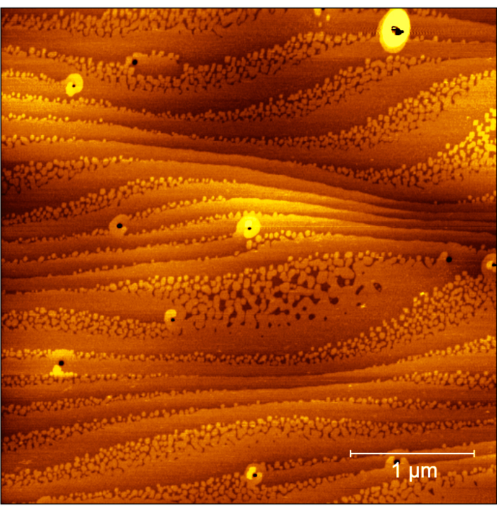
Beautiful Steps
Artist: Kavinraaj Ella Elangovan, student in the Department of Chemical Engineering and Materials Science, University of Minnesota-Twin Cities
NNCI Site: MINIC
Tool: Bruker Nanoscope V Multimode 9 AFM with QNM
The main focus of our research is to develop thin films and heterostructures of perovskite-based quantum materials such as stannates and titanates using a novel hybrid molecular beam epitaxy (MBE) technique. Hybrid MBE gives us high precision control over composition, structure, and strain. The films that we develop are used to better understand and control the interplay between lattice, charge, and spin degrees of freedom I these materials, and their effects on change transport, magnetism, superconductivity, structure, and electronic phase transition. The above image is an atomic force microscopy image of one such thin film (neodymium-doped strontium titanate).
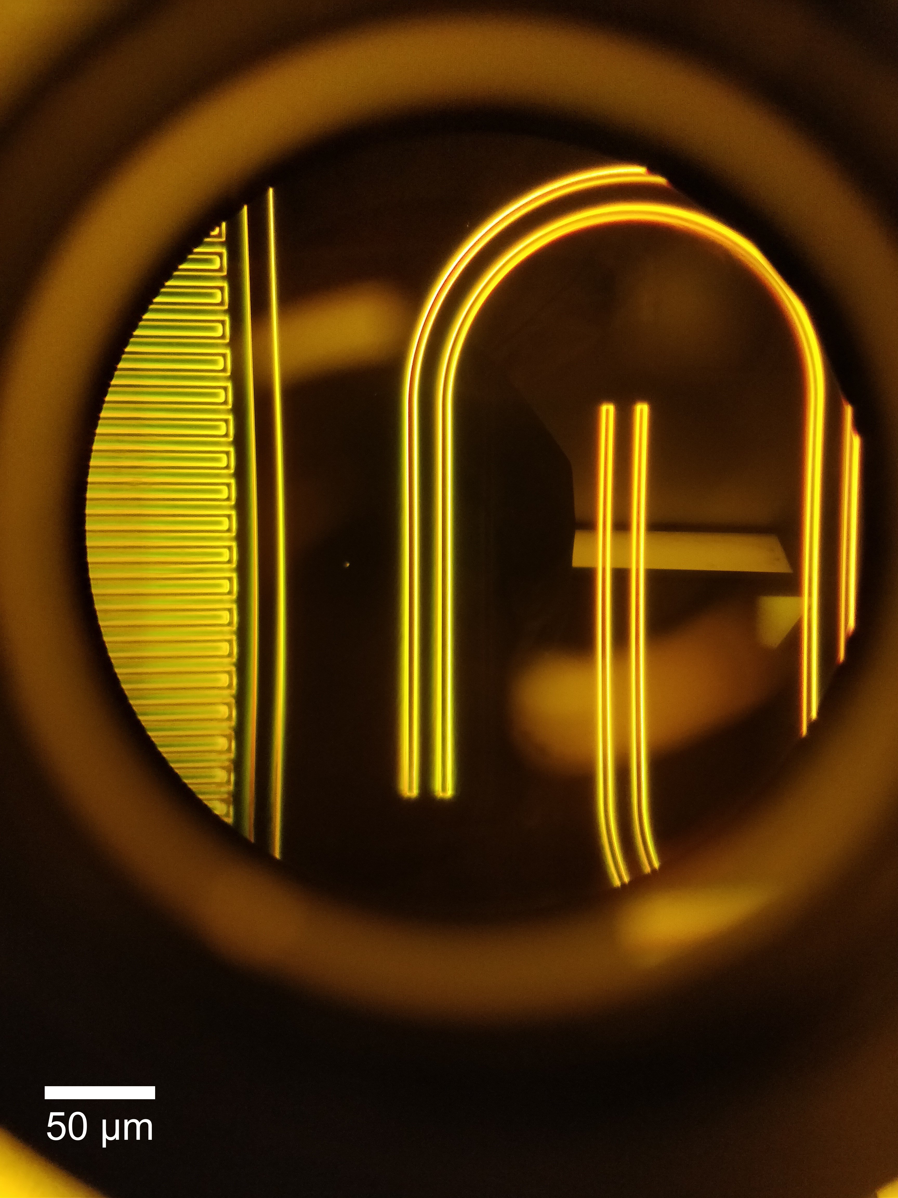
Untitled
Artist: Kevin Multani, graduate student, Stanford University
NNCI Site: nano@Stanford
Tool: Heidelberg2 and optical microscope
A raw darkfield image taken through an optical microscope with my phone in SNF. Seen in the image: (left) mask for an interdigitated capacitor (right) CPW test structures.
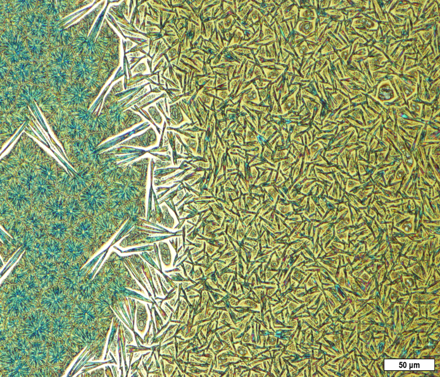
Grain Boundaries
Artists: Calla McCulley, graduate student, under the guidance of Dr. Ananth Dodabalapur, University of Texas at Austin
NNCI Site: TNF
Tool: Olympus optical microscope
Crystallinity controls the overall performance of semiconductors implemented in electronic devices. The left side of this optical image depicts a crystalline film with grain boundaries separating different ordered crystalline domains and the right side of this optical image depicts an amorphous film with needle-shaped crystals. Both types of crystalline structures are beneficial for different electrical device types. This optical image of a methylammonium lead iodide thin film deposited on a zirconium dioxide substrate was acquired in the Microelectronics Research Center at the J.J. Pickle Research Campus.